
iTechGuides is reader-supported. When you buy through links on our site, we may earn an affiliate commission. As an Amazon Associate I earn from qualifying purchases. Learn more
AI can help clinicians find coronary plaque, estimate how much an artery is narrowed, and predict whether a narrowing is reducing blood flow—but it does not perform the scan or diagnose disease by itself. CT, MRI, ultrasound, or a nuclear camera first acquires the images. Software then traces vessels, measures plaque and heart function, detects patterns, and, in some systems, simulates blood flow. A coronary CT angiogram (CCTA) is mainly an anatomic test; stress PET, stress echocardiography, and stress cardiac MRI (CMR) show whether the heart muscle becomes under-supplied or dysfunctional. The most useful result comes from combining the algorithm’s measurements with image quality, symptoms, medical history, and review by a qualified clinician.
What is AI-powered cardiac imaging?
AI-powered cardiac imaging is software-assisted analysis of images produced by established medical scanners. The scanner still supplies the data; the algorithm helps turn thousands of pixels or voxels into measurements that would otherwise take longer to produce manually.
The American Heart Association’s 2024 overview describes applications across echocardiography, cardiac CT, CMR, and nuclear imaging. Examples include outlining the heart’s chambers, calculating ejection fraction, measuring wall motion, quantifying plaque, estimating blood flow, and flagging patterns associated with disease.
Crashes, No Sound, or Screen Glitches?
Random freezes, missing sound and display glitches usually trace back to one bad driver. Find and replace yours safely.Free scan · under a minuteWindows Errors? Fix Them Before They Spread
Repair common Windows errors and clear accumulated junk for a smoother, more stable PC - no reinstall needed.Free scan · no reinstallWhat the software actually does
- Acquisition: CT, MRI, ultrasound, or a nuclear camera records the heart, coronary arteries, or blood-flow response to stress.
- Pre-processing: The system checks and organizes the image series, compensates for motion where possible, and identifies the cardiac phase or relevant anatomy.
- Segmentation and measurement: Algorithms outline chambers and vessels, measure volumes and ejection fraction, and separate calcified and non-calcified plaque from surrounding tissue.
- Risk or physiology modeling: Some systems estimate stenosis, myocardial blood flow, or flow reserve. CT-derived fractional-flow-reserve software builds a three-dimensional model from CCTA data and simulates pressure and velocity.
- Clinical review: A radiologist, cardiologist, or other qualified clinician checks the images, measurements, symptoms, and other tests before making a diagnosis or treatment decision.
That last step matters. The AHA has cautioned that few artificial-intelligence tools have yet shown enough improvement in cardiovascular and stroke care to become widely adopted. AI output is decision support, not an independent medical opinion.
#1 Best Overall
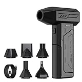
- Powerful Turbo Fan:CAGIWIRU Electric Air Duster have reached speed up to 130000RPM,Can efficient remove dust and debris.With LED light and Three Adjustable Speed to meet with different cleaning tasks.
- With Attached USB C Fasting Charging cable.Charing 3 hours,Enjoy up to 240 minutes of use on the lowest setting, For third speed can use for 25mins.with four charging options to meet with your needs.
- Wide Application: attached 5 different nozzles,making it suitable for a variety of scenes, such as car,pet, pc, keyboards, and other electronic devices. It also serves for office and home clean duster.
- Handy Design & Portable:Cordless air duster electric nozzle is Light weight and small size, design and comfortable to hold and space saving, convenient to carry around. You can put it in your room or inside the car without taking up space.
- Economical &Reusable: Built durable materials with a long-lasting battery,our CAGIWIRU air duster it's a sustainable alternative to disposable air cans.
Can AI see a blocked coronary artery?
It can help show a narrowing and characterize the plaque causing it, especially on CCTA, but a visible narrowing is not automatically a blood-flow problem. CCTA creates an anatomic map of the coronary lumen and artery wall. AI can trace each vessel, calculate the amount and type of plaque, estimate the percentage of narrowing, and highlight segments that deserve expert attention.
Anatomic obstruction versus ischemia
Anatomic stenosis means the artery looks narrower on the image. Ischemia means the narrowing—or another process—is actually limiting oxygen-rich blood reaching the heart muscle. A moderately narrowed segment may not reduce flow at rest, while a less dramatic-looking lesion can become important during exertion or stress.
CT-derived fractional flow reserve (FFRCT) addresses that second question without placing a pressure wire in the artery. Software uses the CCTA-derived three-dimensional coronary model to simulate pressure and flow, producing a physiologic estimate of whether a lesion is likely to reduce blood supply.
A 2023 real-world study of 532 patients reported that AI-assisted CCTA quantified calcification, plaque types, and plaque volume. Combining CCTA with FFRCT could identify physiologically significant disease even when anatomic stenosis was below 50 percent. The investigators also said implementation was feasible but did not replace expert review and clinical correlation.
When invasive angiography is still needed
Catheter-based coronary angiography remains important when anatomy must be confirmed in detail or an intervention such as stent placement is being considered. Noninvasive AI analysis can help determine who needs that next step, but it does not insert a catheter, take a pressure-wire measurement, or treat the blockage.
How does an AI heart scan work from appointment to report?
The exact workflow varies by modality and hospital, but the practical sequence is usually as follows.
Rank #2

- Screen Cleaner is Your Screen’s New Bestfriend! - EVEO is proud to present a screen cleaner that’s perfectly suited for your TV. Meet your new screen cleaner, computer screen cleaner, laptop cleaner, iPad cleaner, Macbook,tv cleaner or any other electronic device. Our screen cleaner spray and wipe along with the included plush microfiber cloth, easily removes dust, fingerprints & other smudges on the screen’s surface. Clean effectively for a streak-free screen
- Gentle on your Screens - Our screen cleaner spray and wipes is compatible to be used even for the most sensitive LCD, LED, CRT, and OLED screens in proper use. Be at ease knowing that this product is compatible with all the major brands: LG, SONY, Samsung, Sharp, iPad, iPhone, Android screens, Kindle, PC monitors - we’ve got you covered! Can be used as LCD screen cleaner, tv cleaner, smart tv screen cleaner, and more
- Premium Super Soft Plush Microfiber Cloth - Included in this efficient screen cleaning kit. This screen cleaner spray and wipe has our signature plush microfiber cloth specially designed to comply with screens and monitors and leave them streak-free. Has been shown to effectively absorbs stubborn stains, zero streaks, and no fiber shedding. Always use this together with our screen cleaner for the best results
- Easy-to-Use Screen Cleaner Kit + microfiber cleaning cloth - The EVEO Screen Cleaning Kit will easily be your new favorite tool when it comes to taking care of the things you love most, quick and easy solution for a streak-free screen. Simply screw on the sprayer gun on the screen cleaner bottle. Spray the cleaning liquid onto the microfiber cloth, then wipe the screen until desired cleanliness & shine is achieved.
- Quality Screen Care Kit Worth Purchasing - Enjoy a convenient Screen Cleaner anytime, anywhere you need it. Your satisfaction is our top priority. We stand behind the quality of our products and that’s why, you shouldn’t be worrying at all. We manufacture the Best Screen Cleaner. and can be used for laptop cleaning kit, laptop screen cleaner, tv screen cleaner for smart tv. MacBook screen cleaner, monitor cleaner or screen cleaner spray for electronic devices with microfiber cleaning cloth
- Clinical question: The clinician chooses an examination based on symptoms, prior results, risk factors, and whether the main question concerns anatomy, ischemia, function, or scar.
- Patient preparation: Staff check factors that can degrade images or change safety decisions, including heart rate and rhythm, body size, kidney function, implanted devices, and claustrophobia. CT examinations may use iodinated contrast; stress and nuclear examinations have their own medication and tracer requirements.
- Image acquisition: The patient lies in the CT, MRI, ultrasound, or nuclear-imaging system. Breath-holding and a steady rhythm can be important for CCTA, while stress studies record the heart at rest and during a controlled increase in workload or blood flow.
- Automated processing: The software receives the scanner’s DICOM image data, segments cardiac structures, measures plaque or function, and may generate a blood-flow simulation or risk score.
- Human verification: A qualified reader reviews the raw images and the overlays or measurements. Poor-quality slices, unusual anatomy, motion, heavy calcium, or artifacts can require correction or manual remeasurement.
- Integrated interpretation: The final report combines the AI findings with symptoms, examination, electrocardiography, laboratory results, prior scans, and clinical judgment.
For example, the FDA describes HeartFlow FFRCT as software that analyzes previously acquired coronary CT DICOM data and supplies computed blood-flow, pressure, and velocity information to support assessment of coronary artery disease. It is intended for qualified clinicians and is not an at-home scan.
Recommended Free Tools
Which cardiac-imaging tests answer which question?
No single modality answers every cardiac question. Choose among them by asking whether the priority is anatomic plaque, flow limitation, muscle function, scar, or a treatment-ready map of the arteries.
| Modality | Main question | What AI can add | Important constraints |
|---|---|---|---|
| Coronary CT angiography (CCTA) | What do the coronary arteries and their plaque look like? | Vessel tracing, stenosis estimates, calcified and non-calcified plaque volume, and automated disease detection. | Image quality can fall with morbid obesity, a high or irregular heart rate, and severe calcification. CT uses ionizing radiation and may require iodinated contrast. |
| CT-derived FFR (FFRCT) | Is a CCTA-visible lesion likely to reduce blood flow? | Three-dimensional pressure and velocity simulation from the CT data. | It depends on a diagnostic-quality CCTA and remains an estimate that must be interpreted clinically. |
| Stress PET | Does myocardial blood flow fall during stress, and how much flow reserve is available? | Quantification of myocardial blood flow and flow reserve, plus automated pattern recognition. | Uses ionizing radiation and a radiotracer; availability and protocol vary. |
| Stress SPECT | Are there stress-related perfusion defects? | Detection and quantification of perfusion patterns. | Uses ionizing radiation and can be affected by attenuation and image artifacts. |
| Stress echocardiography | Does the heart muscle move abnormally when stressed? | Chamber segmentation, wall-motion analysis, and ejection-fraction measurement. | Results depend on acoustic windows and the quality of the ultrasound images. |
| Cardiac MRI (CMR), including stress CMR | What are the heart’s function, wall motion, ischemia, or scar? | Automated chamber volumes, ejection fraction, motion analysis, tissue characterization, and ischemia detection. | Device-safety screening and claustrophobia can limit access; the examination is sensitive to motion and protocol quality. |
| Invasive coronary angiography | What is the detailed arterial anatomy when confirmation or treatment planning is required? | Computer-assisted measurements can support the invasive study, but the procedure itself remains clinician-led. | It is an invasive procedure with catheter, contrast, and procedural risks. |
ACC/AHA criteria describe CCTA as capable of detecting both nonobstructive and obstructive plaque, CT-FFR as a way to assess ischemia, CMR as a test for wall motion, ischemia, and infarction, and PET as a method for quantifying myocardial blood flow and reserve. For appropriate patients with persistent symptoms, the chronic-coronary-disease guideline recommends stress PET or SPECT, CMR, or stress echocardiography to detect ischemia, estimate risk, and guide treatment.
How accurate is AI cardiac imaging?
Published results are encouraging, but accuracy depends on the modality, the patient group, the image quality, and whether the algorithm is being used for measurement or diagnosis. A prospective randomized European Radiology study published in 2024 included 1,801 adults. Fully automated CCTA processing took an average of 121.0 ± 18.6 seconds, compared with 433.5 ± 68.4 seconds for semi-automated processing. Diagnostic accuracy for obstructive coronary artery disease was 94.7 percent with full automation and 94.3 percent with semi-automation, meeting that study’s non-inferiority criterion.
Those figures show that automation can substantially shorten processing without reducing accuracy in that tested setting. They do not mean every AI product is 94.7 percent accurate, nor that the same performance applies to people with different scanners, severe calcium, irregular rhythms, or poor image quality.
Do these 3 things before closing this tab:
1Fix the driver behind crashes, sound loss and screen glitches2Repair Windows errors before they cause bigger problems3Scan for outdated or missing drivers - takes under a minuteA regulated PET example
In 2024, U.S. Food and Drug Administration documentation for the PET tracer Flyrcado reported sensitivity ranging from 74 to 89 percent and specificity from 53 to 70 percent for coronary artery disease defined as at least 50 percent narrowing. The ranges reflect different study conditions and should not be transferred to CCTA, CT-FFR, MRI, or another PET system.
Rank #3

- 【Large size】:Measuring 800 x 300 mm/ 31. 5×12 inches, which is wider and taller than others. Large mouse pad can perfectly fit your keyboard and mouse of any size, and there is also ample space for peripherals such as phones, tablets, etc.
- 【LED backlight mouse pad】:Blade Hawks RGB mouse pad provides 7 static modes and 7 dynamic modes will give you the options you are looking for. Power-off memory function remembers the last lighting mode you selected. The super glow fiber Chroma will give you a colorful gaming setup that you want.
- 【Anti-slip rubber base】:Made from Natural rubber, this gaming mousepad will firmly grip your desktop, allowing you to enjoy the ultimate precision in the games you are most passionate about. It also comes with a texture that enhances this ability even further.
- 【Smooth and waterproof surface】: Micro-textured cloth surface, for extremely precise mouse control and a smooth movement. When liquid splashes on the gaming mouse pad, it forms water droplets and slides down. Will not delay your work or gameplay.
- 【Plug and play】: This mouse and keyboard pad is powered by USB, plug and play, no driver required. One button control light modes. Press one time to change the lighting mode, press twice to change the brightness, press and hold about 3 seconds turn ON/OFF.
What “accurate” should mean to a patient
Ask what the reported metric measures: detecting any plaque, identifying at least 50 percent narrowing, predicting flow-limiting disease, measuring ejection fraction, or finding scar. Sensitivity and specificity describe different errors, and a test’s predictive value also changes with the patient’s underlying likelihood of disease. A clinician can explain how the result changes the next decision rather than treating one percentage as a universal guarantee.
What can make an AI cardiac result unreliable?
Algorithms learn from existing images and labels, so they can inherit limitations in the data used to train and validate them. They may perform less well on populations, scanners, protocols, or disease patterns that were under-represented in development.
- Motion and rhythm: A fast or irregular heart rate can blur coronary arteries and reduce CCTA quality.
- Severe calcification: Calcium blooming can make the artery look more narrowed than it is and complicate plaque segmentation.
- Body size and acoustic windows: Morbid obesity can degrade CT or ultrasound image quality.
- Unusual anatomy or prior treatment: Stents, bypass grafts, congenital variants, and rare disease may require more manual interpretation.
- Incomplete or mismatched data: A flow simulation cannot rescue a CCTA that does not adequately show the vessels, and an automated ejection fraction is only as sound as the chamber boundaries.
- Incidental findings and false alarms: A highlighted region may be clinically insignificant, while a subtle abnormality can be missed. The raw images and patient context remain essential.
The AHA’s position is consistent with these limitations: AI should augment, not replace, cardiovascular expertise. A clinician should be able to inspect the source images, recognize an artifact, and reconcile an algorithmic result with the rest of the case.
Quick wins for a faster PC:
Repair Windows errors before they cause bigger problemsFix Now →Fix the driver behind crashes, sound loss and screen glitchesFind Drivers →What are the safety and patient constraints?
Safety depends more on the imaging modality and the patient than on the word “AI.” Before scheduling a test, the care team considers the following.
- Radiation: CT, PET, and SPECT use ionizing radiation. The ordering clinician weighs the expected diagnostic benefit against exposure and avoids duplicating studies unnecessarily.
- Contrast and kidney function: CCTA and some MRI or angiographic protocols use contrast. Kidney function, prior reactions, medication use, and emergency-care needs affect the choice and preparation.
- Heart rate and rhythm: A high or irregular rate can reduce CCTA quality. The imaging team may need a different protocol or another modality.
- Implanted devices: MRI requires device-specific safety screening and adherence to the scanner manufacturer’s conditions.
- Claustrophobia and ability to remain still: CMR can be difficult for people who cannot tolerate an enclosed scanner or prolonged motion restriction.
- Stress testing: Pharmacologic or exercise stress changes the heart’s workload or blood flow and must be supervised with appropriate monitoring.
Tell the ordering team about pregnancy or possible pregnancy, kidney disease, previous contrast reactions, implanted devices, irregular heartbeat, and difficulty lying flat. They can decide whether the expected information justifies the selected examination or whether another test is safer and more useful.
When is AI-assisted imaging most useful?
AI is especially helpful when it turns a large image set into reproducible measurements that guide a specific decision.
Rank #4

- 【Softer and More Comfortable】Vaydeer wrist rest has unique diamond pattern, which is the combination of softness and aesthetics. The materials of wrist rest are improved into higher quality memory foam and covered with silky smooth lycra. The computer wrist rest makes you as comfortable and cushiony as like rest your wrists on clouds.
- 【Ergonomic Wrist Saver】The wrist rests for keyboard and mouse comes with a 17.32×3.15×0.83 inch keyboard wrist pad and a 5.94×3.15×0.83 inch mouse wrist support. Based on ergonomic design, the unique concave shape is the perfect fit for your wrist joints. The wrist rest pad fits most computer keyboards and laptops, improve hand and wrist posture, release your wrist and arm stress.
- 【Non-Slip Rubber Bottom】Featuring an anti-skid silicone base on the bottom, this wrist keyboard support stays firmly in place on your desk, preventing the padding from sliding around, ensuring stable and consistent wrist support during extended computer sessions.
- 【Better Experience & Pain Relief】Our keyboard arm rest is beneficial to alleviate the soreness caused by direct contact and friction between your arm and a hard desk surface, reducing the risk of wrist fatigue or carpal tunnel. The soft texture of memory foam can evenly distribute the pressure around your wrists and provide good support with just enough give.
- 【Helpful in Multiple Scenarios】Whether you're working, studying, writing, typing, gaming, this keyboard and mouse rest combo is an essential accessory to add comfort and support to your hands and wrists. It’s also a great gift for men, women, family, friend, coworker, gamer, teacher, etc.
Evaluating stable or recurring symptoms
CCTA can show whether plaque is present and whether the lumen appears narrowed. If the anatomic result leaves uncertainty about flow limitation, FFRCT or a stress study may add physiologic information.
Assessing persistent symptoms after an initial test
For appropriate patients whose symptoms continue, guideline-supported stress PET or SPECT, stress CMR, or stress echocardiography can look for inducible ischemia and help estimate risk. The choice depends on prior images, local expertise, radiation considerations, body habitus, kidney function, devices, and patient tolerance.
Planning or triaging invasive care
A detailed noninvasive map can help determine whether catheter angiography is warranted and which lesions need closer evaluation. If intervention is likely, invasive angiography remains the definitive procedural route.
Following heart function over time
Automated chamber contours and ejection-fraction measurements can make serial echocardiography or CMR assessments faster and more consistent. Clinicians still check whether the contours include the correct chambers and whether loading conditions or image quality changed between examinations.
Independent reader supportYour contribution helps us test, update, and keep practical guides available for everyone.How should you read an AI-assisted cardiac-imaging report?
Look for the clinical question and the quality statement before focusing on a single highlighted number.
- Study quality: Note motion, heart-rate limitations, incomplete vessel visualization, or other reasons the reader may have reduced confidence.
- Anatomy: Identify whether the report describes no plaque, nonobstructive plaque, or a narrowing considered obstructive under the report’s criteria.
- Plaque: Check whether calcified, non-calcified, or total plaque burden was measured and whether the finding is in one segment or widespread.
- Physiology: If FFRCT, PET flow, or stress imaging is included, determine whether it indicates a likely flow-limiting problem rather than simply a visible narrowing.
- Function and tissue: For echo or CMR, review ejection fraction, chamber size, regional wall motion, ischemia, and infarct or scar findings.
- Next step: Read the impression and recommendation alongside symptoms and prior tests. An algorithmic flag is not, by itself, a diagnosis or a treatment order.
Is there an AI cardiac-imaging scan for home use?
No. The regulated workflow described here requires a clinical scanner, trained staff, image-data handling, and professional interpretation. Software such as the FDA-described FFRCT application analyzes previously acquired clinical CCTA data; it does not replace the scan and is not a consumer device that can look through the chest at home.
Best Value

- 【No Drill Mounted】Cable management tray can be assembled in as little as 3 minutes or less-simply. Side clips easily clampe the edge of desk, pass through the holes on the sides, and secure excess cables with baskets. Desk cable management supports inward or outward installation to meet your needs in different scenarios, and provides you with great convenience when collecting and organizing wires.
- 【Rubber Pad Protects Desk】Wire organizer under desk built-in soft rubber pads on the clamp protect your desk from scratches, so you will not damage your furniture or office.
- 【Sturdy and Beautiful】Cord management under desk is made of high-quality carbon steel and holds about 15 lbs. The wire basket has 2 holes on each side of the tray to enable your cables to pass freely. Cable baskets are suitable for desk thickness use from 0.4" to 2.4".
- 【Space Saving & Keep Organized】Desk cable management tray holds all your power cords, outlet strips, USB hubs and computer transformers hidden from your feet, and the data cables will not enter the vacuum cleaner to keep your desk clean and tidy. Desk wire organizer is also suitable for use in the kitchen and outdoors.
- 【Reduce Safety Risks】Cable tray under desk keeps all plugs away from your kids, no more worry about tripping. Using 2 holes wire tray saves extra space, helps you easily cabling and wire protection. The mesh design is not easy to accumulate dust, but avoids safety accidents caused by messy wires.
The FDA also recorded a 2024 substantial-equivalence decision for Circle Cardiovascular Imaging’s Cardiac CT Function Software Application. Regulatory clearance or equivalence indicates that a product can be used for its specified medical purpose under its labeling; it does not guarantee that every use, patient, or scanner will produce the same result.
What should you ask before agreeing to an AI-assisted test?
- What decision is this examination intended to answer: plaque, flow limitation, heart function, scar, or treatment planning?
- Will the test use radiation, contrast, a stress drug, or a radioactive tracer?
- What factors in my history—kidney function, heart rhythm, implanted device, body size, or claustrophobia—could affect image quality or safety?
- Will a radiologist or cardiologist review the original images as well as the software measurements?
- If the result is abnormal or inconclusive, what test or follow-up would come next?
Frequently Asked Questions
Can a normal AI-assisted CT scan rule out every heart problem?
No. A coronary CT study mainly evaluates the coronary arteries and related anatomy. It may not explain every cause of chest discomfort, such as a rhythm disorder, valve problem, or disease outside the imaged structures, so persistent or changing symptoms still need clinical assessment.
Does a calcium score show the same thing as an AI coronary CT analysis?
No. A calcium score focuses on calcified plaque. An AI-assisted CCTA can also evaluate the vessel lumen and non-calcified plaque, while a stress or flow-based test addresses whether blood supply becomes inadequate.
What’s actually slowing this PC down?
Pick the symptom - the matching free tool is one click away.
Why can two cardiac-imaging tests disagree?
They may be measuring different endpoints: one test can show anatomy while another measures stress perfusion or wall motion. Differences in timing, scanner quality, heart rate, artifacts, and disease progression can also change the apparent result; clinicians reconcile the studies rather than choosing a number in isolation.
Can AI tell whether chest pain is an emergency?
Imaging software is not an emergency triage system. Sudden or severe chest pain, shortness of breath, fainting, sweating, or other acute warning signs require urgent medical evaluation, where clinicians use symptoms, examination, ECG, blood tests, and imaging as appropriate.
Quick Recap
Product prices and availability are accurate as of the date/time indicated and are subject to change. Any price and availability information displayed on Amazon at the time of purchase will apply.

