
Quick wins for a faster PC:
Repair Windows errors before they cause bigger problemsFix Now →Scan for outdated or missing drivers - takes under a minuteDriver Scan →Clear out junk files and repair common Windows errorsFree Scan →iTechGuides is reader-supported. When you buy through links on our site, we may earn an affiliate commission. As an Amazon Associate I earn from qualifying purchases. Learn more
Most PET/CT artifacts can be separated from true uptake by following a fixed sequence: verify PET-to-CT alignment, inspect the diagnostic CT and attenuation-correction map, compare attenuation-corrected with non-attenuation-corrected PET, identify a matching source such as motion, metal, contrast or truncated anatomy, and then re-register or repeat the acquisition when the defect cannot be corrected. Do not call a suspicious focus malignant until those checks are complete.
Misregistration is identified in SNMMI guidance as the most common source of PET or SPECT/CT artifacts. EANM quality-control guidance separately describes respiratory motion as the most common artifact demonstrated on PET/CT. The distinction matters: respiratory motion is a major cause of misregistration, but other patient movement, CT problems and attenuation-correction errors can produce similar-looking uptake.
What a PET/CT artifact is—and why it can mimic disease
PET measures radiotracer distribution over an emission acquisition, while CT supplies anatomy and an attenuation map used to correct the PET data. The two datasets are acquired at different times and under different physical conditions. If the patient moves, breathes differently, holds a breath for one scan but not the other, or has dense material such as metal or iodinated contrast in the CT field, the attenuation map may no longer describe the anatomy sampled by PET.
Recommended Free Tools
An artifact may therefore appear as a focus of increased or decreased uptake, a distorted lesion, an incorrect standardized uptake value (SUV), or apparent activity in the wrong organ. A focus that aligns with a CT streak, an implant, a contrast column, missing anatomy at the edge of the field of view, or a displaced organ should be treated as potentially technical until proven otherwise.
#1 Best Overall
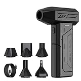
- Powerful Turbo Fan:CAGIWIRU Electric Air Duster have reached speed up to 130000RPM,Can efficient remove dust and debris.With LED light and Three Adjustable Speed to meet with different cleaning tasks.
- With Attached USB C Fasting Charging cable.Charing 3 hours,Enjoy up to 240 minutes of use on the lowest setting, For third speed can use for 25mins.with four charging options to meet with your needs.
- Wide Application: attached 5 different nozzles,making it suitable for a variety of scenes, such as car,pet, pc, keyboards, and other electronic devices. It also serves for office and home clean duster.
- Handy Design & Portable:Cordless air duster electric nozzle is Light weight and small size, design and comfortable to hold and space saving, convenient to carry around. You can put it in your room or inside the car without taking up space.
- Economical &Reusable: Built durable materials with a long-lasting battery,our CAGIWIRU air duster it's a sustainable alternative to disposable air cans.
The step-by-step troubleshooting workflow
- Confirm patient position and overlay alignment. Scroll through axial, coronal and sagittal fused images. Check that the skin, spine, ribs, diaphragm, liver dome and other fixed landmarks occupy the same location on PET and CT. Look especially at the heart, lung bases and upper abdomen, where respiratory or cardiac phase differences are common.
- Review the CT by itself. Determine whether the CT is diagnostic in the region of interest. Look for motion blur, streaks, photon-starvation noise, metal, dense contrast, truncation or anatomy outside the displayed field of view. A poor CT can make the fused image look abnormal even when the PET emission data are acceptable.
- Inspect the attenuation-correction map. The map should follow the patient contour and contain plausible tissue boundaries. Abrupt gaps, streaks, unusually dense regions or a body outline that does not match the CT suggest that attenuation correction may be responsible for the apparent uptake.
- Compare attenuation-corrected and non-attenuation-corrected PET. A focus that disappears or changes substantially on the non-attenuation-corrected series is suspicious for an attenuation-correction artifact. This comparison is specifically recommended when CT truncation or metal artifacts may have propagated into PET.
- Match the focus to a physical cause. Ask whether the uptake follows a CT streak, prosthesis, dental filling, port, contrast-filled vessel or bowel segment, truncated body contour, or a registration mismatch. Correlate in all three planes rather than relying on one fused slice.
- Apply the least disruptive correction first. If both PET and CT are individually usable, software re-registration can correct a rigid positional offset. If motion has degraded either the emission or transmission dataset, re-registration may not recover the lost information; a repeat acquisition may be needed when the result would change management.
- Document the effect on interpretation and quantification. State the suspected cause and whether it affects only the visual impression, the SUV, or both. A technically compromised SUV should not be compared with prior or follow-up values as if it were measured under equivalent conditions.
Motion and PET/CT misregistration
How it appears
Patient movement between the PET emission scan and the CT transmission scan shifts organs or the entire body contour. On fused images, uptake may appear adjacent to rather than within a lesion, cross an organ boundary, or be assigned to the wrong structure. The diaphragm, liver dome, lung bases and heart are particularly vulnerable. A rigid shift can affect a large region; smaller movements can create a local mismatch around the shoulders, arms or pelvis.
How to troubleshoot it
- Recheck the external contour, spine and pelvic bones before judging a soft-tissue focus.
- Review PET and CT in separate windows to establish whether each dataset is internally coherent.
- Use the scanner’s validated software re-registration tools when the problem is a simple positional offset and the PET and CT images remain diagnostically sound.
- After re-registration, reassess the focus on the corrected fusion and on non-attenuation-corrected PET. Do not assume that a visually improved overlay restores an accurate SUV if the original acquisition was degraded.
If movement occurred during the PET emission scan, counts from different positions may be blended and cannot always be repaired by shifting the CT. If motion occurred during CT, the attenuation map itself may be unreliable. In either case, consider repeat imaging when the abnormality is clinically important and no correction produces a trustworthy result.
Respiratory mismatch
Why the lung–liver and heart–diaphragm interfaces are difficult
Respiratory motion is described in EANM quality-control guidance as the most common artifact demonstrated on PET/CT. PET usually samples several minutes of breathing, whereas a diagnostic CT may be obtained during a brief deep-inspiration breath-hold or during quiet breathing. The liver, spleen, diaphragm, heart and lower lungs can therefore occupy different positions in the two datasets.
Do these 3 things before closing this tab:
1Clear out junk files and repair common Windows errors2Scan for outdated or missing drivers - takes under a minute3Repair Windows errors before they cause bigger problemsRank #2

- Screen Cleaner is Your Screen’s New Bestfriend! - EVEO is proud to present a screen cleaner that’s perfectly suited for your TV. Meet your new screen cleaner, computer screen cleaner, laptop cleaner, iPad cleaner, Macbook,tv cleaner or any other electronic device. Our screen cleaner spray and wipe along with the included plush microfiber cloth, easily removes dust, fingerprints & other smudges on the screen’s surface. Clean effectively for a streak-free screen
- Gentle on your Screens - Our screen cleaner spray and wipes is compatible to be used even for the most sensitive LCD, LED, CRT, and OLED screens in proper use. Be at ease knowing that this product is compatible with all the major brands: LG, SONY, Samsung, Sharp, iPad, iPhone, Android screens, Kindle, PC monitors - we’ve got you covered! Can be used as LCD screen cleaner, tv cleaner, smart tv screen cleaner, and more
- Premium Super Soft Plush Microfiber Cloth - Included in this efficient screen cleaning kit. This screen cleaner spray and wipe has our signature plush microfiber cloth specially designed to comply with screens and monitors and leave them streak-free. Has been shown to effectively absorbs stubborn stains, zero streaks, and no fiber shedding. Always use this together with our screen cleaner for the best results
- Easy-to-Use Screen Cleaner Kit + microfiber cleaning cloth - The EVEO Screen Cleaning Kit will easily be your new favorite tool when it comes to taking care of the things you love most, quick and easy solution for a streak-free screen. Simply screw on the sprayer gun on the screen cleaner bottle. Spray the cleaning liquid onto the microfiber cloth, then wipe the screen until desired cleanliness & shine is achieved.
- Quality Screen Care Kit Worth Purchasing - Enjoy a convenient Screen Cleaner anytime, anywhere you need it. Your satisfaction is our top priority. We stand behind the quality of our products and that’s why, you shouldn’t be worrying at all. We manufacture the Best Screen Cleaner. and can be used for laptop cleaning kit, laptop screen cleaner, tv screen cleaner for smart tv. MacBook screen cleaner, monitor cleaner or screen cleaner spray for electronic devices with microfiber cleaning cloth
A deep-inspiration diagnostic CT substituted for a normal-breathing attenuation CT can create unacceptable misregistration. The resulting apparent uptake may be placed in the wrong lobe, appear as a thin band at the diaphragm, or be split between liver and lung.
Practical checks and corrections
- Inspect the lung–liver interface and the heart–diaphragm interface in all planes.
- Determine whether the CT was acquired with breath-hold instructions that differ from those used for the attenuation scan or PET acquisition.
- Use a respiratory protocol matched to the PET acquisition when available. Avoid inconsistent instructions between the transmission CT and any diagnostic breath-hold CT.
- Use respiratory-gated or motion-matched reconstruction only when supported by the local scanner protocol and the clinical question; gating changes acquisition and reconstruction characteristics and should not be assumed to be interchangeable with routine whole-body PET/CT.
- Re-register only when the mismatch is a correctable rigid displacement. A continuously moving diaphragm cannot be made accurate by a simple translation.
When a focus lies at the moving interface and remains indeterminate after review, describe the limitation and correlate with diagnostic CT, MRI, ultrasound or follow-up imaging as clinically appropriate rather than assigning a precise SUV to the mismatched voxel.
Metal, implants and dental hardware
Mechanism and visual clues
Ports, prostheses, orthopedic hardware, spinal fixation, pacemakers and dental restorations can cause severe CT streaking. The CT reconstruction may assign incorrect attenuation coefficients around the object. CT-based attenuation correction can then create falsely high or falsely low PET activity near the hardware, impairing both lesion detection and quantification.
Rank #3

- 【Large size】:Measuring 800 x 300 mm/ 31. 5×12 inches, which is wider and taller than others. Large mouse pad can perfectly fit your keyboard and mouse of any size, and there is also ample space for peripherals such as phones, tablets, etc.
- 【LED backlight mouse pad】:Blade Hawks RGB mouse pad provides 7 static modes and 7 dynamic modes will give you the options you are looking for. Power-off memory function remembers the last lighting mode you selected. The super glow fiber Chroma will give you a colorful gaming setup that you want.
- 【Anti-slip rubber base】:Made from Natural rubber, this gaming mousepad will firmly grip your desktop, allowing you to enjoy the ultimate precision in the games you are most passionate about. It also comes with a texture that enhances this ability even further.
- 【Smooth and waterproof surface】: Micro-textured cloth surface, for extremely precise mouse control and a smooth movement. When liquid splashes on the gaming mouse pad, it forms water droplets and slides down. Will not delay your work or gameplay.
- 【Plug and play】: This mouse and keyboard pad is powered by USB, plug and play, no driver required. One button control light modes. Press one time to change the lighting mode, press twice to change the brightness, press and hold about 3 seconds turn ON/OFF.
Look for a streak or dark band on CT that continues into the attenuation map and overlies the PET focus. Dental hardware commonly affects the head and neck; shoulder prostheses and spinal hardware can affect adjacent chest, neck or abdominal structures. The artifact may be asymmetric and may not follow a physiologic distribution.
Correction sequence
- Review the original CT and attenuation map side by side with fused PET.
- Use a validated metal-artifact-reduction reconstruction when the scanner provides one. Treat it as a reconstruction aid, not proof that the underlying PET activity is real.
- Compare with non-attenuation-corrected PET. Disappearance or major reduction of the focus implicates attenuation overcorrection.
- Assess whether the area is still interpretable on the CT itself. If streaking obscures anatomy, avoid an exact SUV-based conclusion.
- If the focus is clinically decisive and no reconstruction resolves it, consider a targeted repeat acquisition or another modality.
Iodinated intravenous or oral contrast
Dense iodinated contrast can be interpreted by CT-based attenuation correction as excessive material. If contrast has moved between the CT and PET portions of the examination, the attenuation map may no longer match the tracer distribution. This can generate false-positive activity, particularly along a contrast-filled vessel, collecting system or bowel segment.
Trace the apparent focus back to the CT contrast column and note whether the location is physiologic for an injected bolus or oral contrast. Then inspect non-attenuation-corrected PET. A focus present only after attenuation correction should not be called a lesion without corroboration. Contrast can also produce a genuine anatomic finding on CT, so the CT appearance and the clinical question must be considered separately from the PET artifact.
Rank #4

- 【Softer and More Comfortable】Vaydeer wrist rest has unique diamond pattern, which is the combination of softness and aesthetics. The materials of wrist rest are improved into higher quality memory foam and covered with silky smooth lycra. The computer wrist rest makes you as comfortable and cushiony as like rest your wrists on clouds.
- 【Ergonomic Wrist Saver】The wrist rests for keyboard and mouse comes with a 17.32×3.15×0.83 inch keyboard wrist pad and a 5.94×3.15×0.83 inch mouse wrist support. Based on ergonomic design, the unique concave shape is the perfect fit for your wrist joints. The wrist rest pad fits most computer keyboards and laptops, improve hand and wrist posture, release your wrist and arm stress.
- 【Non-Slip Rubber Bottom】Featuring an anti-skid silicone base on the bottom, this wrist keyboard support stays firmly in place on your desk, preventing the padding from sliding around, ensuring stable and consistent wrist support during extended computer sessions.
- 【Better Experience & Pain Relief】Our keyboard arm rest is beneficial to alleviate the soreness caused by direct contact and friction between your arm and a hard desk surface, reducing the risk of wrist fatigue or carpal tunnel. The soft texture of memory foam can evenly distribute the pressure around your wrists and provide good support with just enough give.
- 【Helpful in Multiple Scenarios】Whether you're working, studying, writing, typing, gaming, this keyboard and mouse rest combo is an essential accessory to add comfort and support to your hands and wrists. It’s also a great gift for men, women, family, friend, coworker, gamer, teacher, etc.
Truncation and field-of-view errors
Recognizing missing anatomy
If part of the patient lies outside the CT field of view, the attenuation map may contain an incomplete body contour. The PET emission data can extend beyond that contour, so correction factors at the edge become inaccurate. Truncation commonly affects broad shoulders, arms alongside the body, large body habitus or a patient who is not centered in the gantry.
Fixes
- Check whether the body contour is cut off on CT or the attenuation map while activity remains visible on PET.
- Confirm that the patient was centered and positioned within the available CT field of view.
- Use the scanner’s extended-field-of-view capability when available and validated for the examination.
- Inspect non-attenuation-corrected PET and avoid quantitative interpretation in the truncated region unless the correction has been verified.
- If the missing anatomy contains a clinically important focus and no reliable reconstruction is possible, discuss repeat imaging with appropriate positioning or an alternative modality.
CT motion, noise and reconstruction artifacts
CT motion blur, low photon counts and reconstruction abnormalities can corrupt the attenuation map even when the patient appears aligned overall. A noisy or streaked CT may create false boundaries on the fused display, while a technically acceptable PET series can be misinterpreted because its correction map is defective.
Review CT quality independently from PET quality. Determine whether the defect is confined to CT, confined to PET, or propagated into both through attenuation correction. Then compare corrected and non-corrected PET. If the PET focus is stable on non-corrected images and the CT defect has no anatomic correlate, the finding is less likely to be an attenuation artifact; if it appears only on corrected PET adjacent to the CT defect, technical over- or under-correction is more likely.
Best Value

- 【No Drill Mounted】Cable management tray can be assembled in as little as 3 minutes or less-simply. Side clips easily clampe the edge of desk, pass through the holes on the sides, and secure excess cables with baskets. Desk cable management supports inward or outward installation to meet your needs in different scenarios, and provides you with great convenience when collecting and organizing wires.
- 【Rubber Pad Protects Desk】Wire organizer under desk built-in soft rubber pads on the clamp protect your desk from scratches, so you will not damage your furniture or office.
- 【Sturdy and Beautiful】Cord management under desk is made of high-quality carbon steel and holds about 15 lbs. The wire basket has 2 holes on each side of the tray to enable your cables to pass freely. Cable baskets are suitable for desk thickness use from 0.4" to 2.4".
- 【Space Saving & Keep Organized】Desk cable management tray holds all your power cords, outlet strips, USB hubs and computer transformers hidden from your feet, and the data cables will not enter the vacuum cleaner to keep your desk clean and tidy. Desk wire organizer is also suitable for use in the kitchen and outdoors.
- 【Reduce Safety Risks】Cable tray under desk keeps all plugs away from your kids, no more worry about tripping. Using 2 holes wire tray saves extra space, helps you easily cabling and wire protection. The mesh design is not easy to accumulate dust, but avoids safety accidents caused by messy wires.
How to distinguish an artifact from a real lesion
| Observation | Interpretation to consider | Next check |
|---|---|---|
| Focus disappears on non-attenuation-corrected PET | Attenuation-correction artifact is likely, especially with metal, contrast or truncation | Inspect CT and attenuation map for the matching cause; do not rely on SUV |
| Focus shifts across an organ boundary on fused images | Registration or respiratory mismatch | Compare fixed landmarks and review the lung–liver or heart–diaphragm interface |
| Focus follows a CT streak from hardware | Metal-induced over- or under-correction | Use validated metal-artifact reduction and confirm on non-corrected PET |
| Activity is at the edge of a cut-off body contour | CT truncation or field-of-view error | Check extended-FOV reconstruction and patient centering |
| Focus remains on both corrected and non-corrected PET with a matching CT lesion | Technical artifact is less likely, although motion can still affect localization | Assess lesion morphology, physiologic distribution and clinical context |
| CT is blurred or noisy while PET appears otherwise uniform | CT motion or insufficient CT count density | Review attenuation map and determine whether a repeat CT or PET/CT is needed |
Persistence on non-attenuation-corrected PET does not by itself prove malignancy; physiologic uptake, injection-related effects and true benign disease remain possible. It simply makes a pure attenuation-correction artifact less likely.
Choosing re-registration, reconstruction or a repeat scan
Use the following decision framework rather than a fixed SUV or displacement threshold. The cited guidance does not establish one universal numeric threshold for repeating an artifact-affected PET/CT, and no single vendor-neutral metal-artifact-reduction setting applies to every scanner. Tracer, body region, local software, acquisition protocol and the clinical question all affect the decision.
| Situation | Preferred action | Why | Main limitation |
|---|---|---|---|
| Simple rigid shift; PET and CT individually diagnostic | Software re-registration, then reassess all planes | Can restore anatomic localization without reacquiring data | Cannot repair motion-blurred emission or transmission data |
| Respiratory mismatch at diaphragm or heart | Use matched respiratory protocol or gated reconstruction when available; interpret cautiously | Addresses phase differences between scans | Continuous motion may remain and SUV comparability can change |
| Metal streak with available validated MAR | Review MAR and standard reconstructions; confirm on non-corrected PET | May reduce incorrect attenuation coefficients | Residual streaks and altered image texture can persist |
| Truncated anatomy with extended-FOV capability | Reconstruct with validated extended FOV and inspect map | Restores a more complete attenuation contour | Accuracy at the extreme edge may remain limited |
| Motion or CT defect that is not correctable and clinically important | Consider repeat acquisition or another modality | New data may be required for a reliable answer | Additional radiation, time, cost and patient burden |
Reporting an artifact-affected examination
A useful report separates what is observed from what is technically uncertain. Include:
- the suspected source: patient movement, respiratory mismatch, metal, contrast, truncation, CT noise or another reconstruction problem;
- the anatomic region affected and whether the PET-to-CT overlay is displaced;
- whether the focus is present on non-attenuation-corrected PET;
- whether the CT itself shows a corresponding lesion or only an artifact;
- whether qualitative interpretation is possible and whether SUV quantification is unreliable;
- any re-registration or reconstruction performed and its effect;
- whether the limitation is important enough to justify repeat imaging or correlation with another modality.
For example, a report can state that uptake at the hepatic dome is inseparable from respiratory misregistration, is reduced on non-attenuation-corrected images, and should not be quantified; or that focal uptake adjacent to a hip prosthesis corresponds to CT streaking and is not confirmed on non-corrected PET. Such wording tells the referring clinician exactly what is uncertain and why.
Quick Recap
Common troubleshooting mistakes
- Reading the fused image first. Fusion can make a misplaced focus look anatomically convincing. Review standalone CT and PET before accepting the overlay.
- Trusting the SUV without checking correction. Metal, contrast and truncation can change attenuation factors and produce misleading numbers.
- Using only one plane. A streak or mismatch that is subtle axially may be obvious coronally or sagittally.
- Assuming re-registration fixes motion. Translation can correct a rigid offset, not blurred or continuously moving data.
- Calling every non-corrected focus an artifact. Persistence on non-corrected PET reduces the likelihood of an attenuation artifact but does not establish the diagnosis.
- Repeating automatically. There is no universal repeat threshold. Balance technical irreparability, clinical importance, patient risk and whether another modality can answer the question.
A concise checklist for daily reading
- Is the patient contour and skeletal anatomy aligned?
- Are the diaphragm, liver dome and heart in the same phase and position?
- Is the CT diagnostic, or is there motion, noise, streaking or truncation?
- Does the attenuation map match the CT and body contour?
- Does the focus persist on non-attenuation-corrected PET?
- Does it follow metal, contrast, a CT streak or a missing field of view?
- Can validated re-registration or reconstruction correct the problem?
- Is the remaining uncertainty acceptable, or would repeat imaging change management?
Product prices and availability are accurate as of the date/time indicated and are subject to change. Any price and availability information displayed on Amazon at the time of purchase will apply.

