
Free tools Windows power users keep installed
One-click scans. No signup required.
A camera system built to recognize loose pastries at checkout became the starting point for a medical image-analysis project. BRAIN Co.’s Cyto-AiSCAN is described as a tool to support cytology review—not an autonomous cancer diagnostic—and a pathologist remains responsible for the final diagnosis.
How BakeryScan recognizes pastries
BRAIN Co., a Japanese image-recognition company, developed BakeryScan for bakeries where products are sold loose and manual identification can slow checkout. A barcode is not practical for every unwrapped pastry, and visually similar items can be difficult to distinguish quickly.
- A camera captures an image of the products on a tray or counter.
- The software analyzes visible characteristics such as shape, color, size, and surface appearance.
- It suggests a likely product and price. If identification is uncertain, it presents candidates for a cashier to choose from.
- The cashier can correct a suggestion, providing feedback to the system.
This human-in-the-loop design matters: the tool can help with routine recognition without being expected to get every item right on its own. BRAIN describes BakeryScan and its image-recognition work on its official site; The New Yorker’s account also describes the bakery checkout problem and the system’s development.
How a bakery system led to a medical project
In early 2017, according to reporting by The New Yorker, a physician associated with Kyoto’s Louis Pasteur Center for Medical Research saw a television segment about BakeryScan. The physician recognized a broad visual analogy: software that distinguishes objects in an image might be adapted to help examine cells under a microscope. The physician contacted BRAIN, and the company began work with medical researchers on a pathology application.
Recommended Free Tools
#1 Best Overall
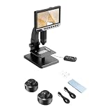
- The ergonomic ABS body microscope combines laboratory grade optics with touch display controls, making professional levels inspection accessible for STEM education and industrial maintenance applications
- Equipped with built in adjustable LED lighting and multimedia captures capabilities, this portable microscope delivers clear visuals across lighting conditions while enabling photo/video documentation
- Designed for electronics technicians, biology enthusiasts, and precise requiring clear magnification for circuit board repairs, analysis, or miniature assembly tasks
- Optimize workshop efficiency by examining solder joints or captures high resolution footage of microorganisms during field research and educational demonstrations
- Explore the microscopic world with our 7 inch LCD digital microscope, featuring high resolution imaging and adjustable magnification for detailed observation of cells, and PCB components
The analogy is about the image-recognition task, not biology. A pastry is not a cancer cell, and the bakery checkout system did not simply become a cancer detector. Medical use required a different imaging environment, relevant cell data, evaluation, and clinical oversight. Accounts of the collaboration and technology’s development are also available from J-Net21 and Digital Garage Laboratory.
What Cyto-AiSCAN is designed to do
Cyto-AiSCAN is described as an AI-based cytology diagnostic-support system. In cytology, specialists examine cells to assess whether they appear normal or abnormal. Company-linked and regional medical-technology descriptions say the system analyzes cellular appearance and can quantify features such as size and atypia, then help draw a reviewer’s attention to cells or regions that merit examination.
Rank #2

- Intended Use: The 120 microscope slides with specimens suitable for science class, homeschool & biology curriculum, various samples covering different topics like biology, botany, zoology, histology, microbiology, cells.
- Microscope Slide Kit Preparation: The Lab-Grade microscope slides with case made with high-quality glass materials and techniques. Well-prepared slides provide clearer images and durable enough for repeated use, complement STEM biology science education
- Labeling & Identification Guide: Microscope glass slides Color labels classified by course to facilitate use, clearly labeled with specimen details like name, magnification, include an identification chart guide which made this microscope accessories kit easy to dig deeper than just knowing the name of the specimen
- Glass Slides Storage & Protection: Comes with a sturdy, organized case to protect the slides for microscope from damage and dust, compatible with most microscope's stage size and objectives, such as compound, stereo, digital, and pocket microscopes
- Prepared Slides Age Appropriateness: The childrens microscope slides contain a variety of samples such as plant, animal, microbiology, tissues, cells, bacteria, fungi, histology, for teaching, studying various biological specimens, ideal microscope slides for kids ages 11+ and adults
The Kobe Biomedical Innovation Cluster lists cervical and bladder cancer examinations among the system’s applications. Descriptions refer to microscopy or whole-slide imaging workflows, but do not establish that the system handles every cancer type or every kind of specimen. See the cluster’s company profile and the Expo-related system description.
What the AI does—and what it does not
Cyto-AiSCAN is presented as assistance for a medical professional, not as a replacement for one. It may help organize review, measure visual features, or flag areas for closer attention. The pathologist interprets the findings in context and makes the final diagnosis, as the Expo-related description states.
Rank #3
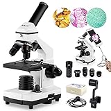
- [High Magnification] WF25X eyepiece alone or with 2X lens, cooperate 4x 10x 40x objective lenses, biological microscopes offers 6 magnification settings, 100X, 250X, 1000X, 200X, 500X, 2000X.
- [Coarse and Fine Focus] Monocular Microscopes with solid metal frame and coarse/fine focusing knobs, ensures detail and precise focus adjustment, crate a image more clearer and sharper.
- [Dual Illumination System] Top and bottom LED simultaneously brightfield, a 0.65 NA single-lens condenser with disc diaphragm, provide high-resolution color-corrected images.
- [Complete Accessories] It comes with phone adapter, wire shutter, 10 operating accessories, 15pcs microscope slides set, carrying bag. Meet all your biological observation need.
- [Practical Educational Tool] This 100X-2000X biological microscope is designed to help students/kids/adults/beginner/amateur scientist/hobbyists exploring the fantastic world of the life. It is suitable for school,laboratory or home teaching, one-on-one instruction.
That distinction is especially important because image patterns are not diagnoses by themselves. A highlighted cell or a measurement can support an expert’s assessment, but it does not independently establish malignancy. This is a research and clinical-support story, not a consumer tool for patients to use to check whether they have cancer.
What the reported accuracy figures mean
Public accounts cite several high-90-percent figures: one retelling gives 97% or higher for BakeryScan identification, some accounts cite 98% for early cancer-cell identification tests, and later secondary descriptions mention 99% for Cyto-AiSCAN. These are reported figures, not interchangeable measures of clinical performance. The figures appear in sources including Analytics Vidhya, The New Yorker, and BRAIN- or cluster-linked descriptions (BRAIN; Kobe Biomedical Innovation Cluster).
Rank #4

- 【Professional Magnification & Clarity】Microscope is equipped with WF25X and 10X eyepieces,4X, 10X, and 100X objective lenses, offering 6 magnification levels (40X to 2500X). The precision-engineered optics provide exceptional visual quality and crystal-clear images, perfect for advanced laboratory work
- 【Dual Illumination System】Featuring a dual LED light source with incident and transmitted LED illumination, you can view opaque specimens with the upper LED or translucent specimens with the lower LED. An adjustable dimmer switch allows you to customise the light intensity for optimal viewing.
- 【Five-Color Filter Wheel】The built-in rotating disc beneath the stage includes five different colored filters (yellow, red, blue, green, and purple), eliminating the need for staining slides. These filters enhance observation of transparent specimens, enabling the capture of high-quality images with ease
- 【User-Friendly Design】Designed with a 360° rotating head and a 45° inclined viewing angle, this binocular microscope minimizes physical strain and facilitates shared viewing, making it ideal for educational settings and professional environments. The adjustable interpupillary distance allows for personalized adjustments, ensuring the best possible observation experience
- 【Dual Control Focusing System】Microscope features both coarse and fine focus knobs. The coarse focus knob quickly locates the image on the stage, while the fine focus knob maintains precise clarity, ensuring details are sharp and well-defined. Enjoy a clear and focused view of your specimens with ease
The available public descriptions do not consistently specify the sample size, cancer type, test design, comparison standard, or whether a percentage means accuracy, sensitivity, specificity, or another metric. They also do not make clear whether a result applies to individual cells, selected image fields, or whole slides. Without those details, a high percentage cannot tell a reader how often the system would miss disease, flag benign cells, or perform on a different hospital’s slides. It should not be restated as “the AI diagnoses cancer with 98% accuracy.”
How the system analyzes images
The technology belongs to the broader field of computer vision and image recognition. Public descriptions refer to visual features including shape, size, color, cellular or nuclear appearance, and atypia. The system detects patterns in images; it does not understand cancer in the way a pathologist understands disease biology.
Best Value
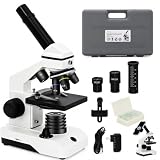
- 🔬 40X–2000X MAGNIFICATION & PREMIUM OPTICS – Two wide-field eyepieces (10× & 25×) plus 4×/10×/40× objectives and a 2X additional lens deliver crystal-clear views of cells, insects, crystals, and more.
- 💼 ALL-IN-ONE LAB KIT – Comes ready to explore with prepared slides, blank slides, tweezers, dropper, and 50 lens-cleaning tissues.
- 📷 SMARTPHONE ADAPTER FOR EASY SHARING – Snap photos or record video right through the eyepiece and share your discoveries instantly on social media or in class projects.
- 🎒 PORTABLE SUITCASE & RUGGED METAL BUILD – Hard-shell case stores every component safely, while the all-metal frame and quality mechanics stand up to classroom and field use.
- 🌟 STEM-FRIENDLY & VERSATILE FOR ALL AGES – Simple enough for beginners, powerful enough for hobbyists and professionals—perfect for home labs, classrooms, and on-the-go science adventures.
Descriptions of its algorithmic approach are not fully consistent. Analytics Vidhya describes BakeryScan in terms of deep learning, while a J-Net21 account contrasts BRAIN’s approach with conventional deep-learning systems and emphasizes image features, expert feedback, and the possibility of working with less data. The public material does not disclose enough technical detail to say definitively whether Cyto-AiSCAN uses or does not use deep learning. It is safer to describe it as AI-based image analysis whose precise architecture is not fully public.
Independent reader supportYour contribution helps us test, update, and keep practical guides available for everyone.Where the technology stood in public accounts
The New Yorker and Analytics Vidhya reported testing or evaluation at major hospitals in Kyoto and Kobe. More recent company-linked descriptions continue to present Cyto-AiSCAN as a development or diagnostic-support system. The cited public sources do not establish broad routine deployment, autonomous clinical use, or a particular regulatory clearance. Anyone evaluating it for clinical procurement would need current details from the company and relevant regulators about intended use, validation, approval status, and deployment conditions.
Why this approach is promising—and what still needs proving
Potential value in review
- Triage: Flagging suspicious-looking cells could help a specialist decide where to look more closely.
- Throughput: Automated image review could help manage large volumes of cells or slides, depending on workflow and validated performance.
- Consistent measurements: Software can quantify visual features in a repeatable way, giving a pathologist additional information to consider.
- Human review: The bakery system’s correction loop illustrates a practical design principle: software can assist while leaving uncertain decisions to people. It does not, by itself, establish how Cyto-AiSCAN’s clinical feedback process works.
Risks and evidence questions
- False negatives: Missing an abnormal cell could delay further review or diagnosis.
- False positives: Flagging benign cells may add unnecessary review or follow-up.
- Differences between settings: Staining, scanners, slide preparation, patient populations, and cancer subtypes can differ between institutions; performance on one dataset may not transfer to another.
- Image quality: Blur, artifacts, debris, overlapping cells, or poor preparation may make reliable recognition harder.
- Misleading overall accuracy: When abnormal cells are uncommon, a high overall score may conceal poor detection of the cases that matter most.
- Clinical readiness: A promising image-analysis result does not by itself show improved patient outcomes or establish that a system is ready for routine use.
- Operational needs: Clinical use also raises questions about workflow integration, staff training, cybersecurity, data handling, maintenance, and regulatory requirements.
A careful evaluation would ask which cells and cancer types were tested; how many patients and slides were included; whether the test used previously unseen slides; and whether it was independently validated across hospitals and scanners. It would also report sensitivity, specificity, predictive values, uncertainty handling, and disagreements with pathologists—not just a single accuracy percentage.
What the bakery-to-cancer story really shows
BRAIN’s broader image-recognition work has been described in other settings, including pill identification, food self-checkout, and food-label verification; the company’s site and FOOMA Japan exhibitor profile describe related applications. Those examples show that visual-recognition methods can be adapted to different tasks, not that one system carries the same performance or clinical value everywhere.
The Tool Desk
Outbyte Driver Updater FREEScan for outdated or missing drivers - takes under a minuteDriver Scan →Outbyte PC Repair FREERepair Windows errors before they cause bigger problemsFix Now →The notable connection is a transfer of image-analysis ideas: recognizing products gave BRAIN a starting point for work with medical images, where specialists and clinical validation remain essential. BakeryScan made checkout recognition a practical problem; Cyto-AiSCAN applies related computer-vision thinking to cytology support. The result is an intriguing bridge between industries, not evidence that a bakery machine independently diagnoses cancer.
Quick Recap
Product prices and availability are accurate as of the date/time indicated and are subject to change. Any price and availability information displayed on Amazon at the time of purchase will apply.

