
Do these 3 things before closing this tab:
1Fix the driver behind crashes, sound loss and screen glitches2Clear out junk files and repair common Windows errors3Scan for outdated or missing drivers - takes under a minuteiTechGuides is reader-supported. When you buy through links on our site, we may earn an affiliate commission. As an Amazon Associate I earn from qualifying purchases. Learn more
Short answer: fMRI is usually the better tool for asking where visual information is represented across brain patterns, while EEG is usually better for asking when that information appears and changes. They measure different physiological signals, so their decoding results are complementary rather than interchangeable. In either case, a successful decoder shows that recorded activity contains information that distinguishes the tested conditions; it does not amount to unrestricted mind reading or prove that a particular region performs the computation identified by the model.
What fMRI and EEG actually measure
fMRI: an indirect, spatially detailed signal
Functional magnetic resonance imaging (fMRI) tracks changes in blood oxygenation and flow that accompany neural activity. The resulting blood-oxygenation-linked signal is an indirect marker: it reflects vascular consequences of neural processing rather than electrical firing itself. Researchers can analyze activity in many small volume elements (voxels) and ask whether the pattern across those voxels differs between visual conditions.
That spatial sampling makes fMRI useful for multivoxel pattern analysis. Instead of asking only whether one area is more active, a study can test whether distributed patterns contain information about a line orientation, object category, scene, attended item or remembered image. Specialized ultra-high-field systems can produce submillimeter imaging in particular research settings, as reviewed in Ultra-High Field Imaging of Human Visual Cognition (2023), but higher spatial detail does not remove the method’s relatively slow hemodynamic response and should not be treated as a property of every routine scan.
EEG: a fast measurement of summed electrical activity
Electroencephalography (EEG) records voltage differences at electrodes placed on the scalp. These potentials reflect the summed, synchronized activity of large populations of neurons, filtered through the tissues between cortex and sensors. EEG therefore provides millisecond-scale information about changing electrical patterns, but each electrode mixes activity from multiple sources.
#1 Best Overall
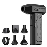
- Powerful Turbo Fan:CAGIWIRU Electric Air Duster have reached speed up to 130000RPM,Can efficient remove dust and debris.With LED light and Three Adjustable Speed to meet with different cleaning tasks.
- With Attached USB C Fasting Charging cable.Charing 3 hours,Enjoy up to 240 minutes of use on the lowest setting, For third speed can use for 25mins.with four charging options to meet with your needs.
- Wide Application: attached 5 different nozzles,making it suitable for a variety of scenes, such as car,pet, pc, keyboards, and other electronic devices. It also serves for office and home clean duster.
- Handy Design & Portable:Cordless air duster electric nozzle is Light weight and small size, design and comfortable to hold and space saving, convenient to carry around. You can put it in your room or inside the car without taking up space.
- Economical &Reusable: Built durable materials with a long-lasting battery,our CAGIWIRU air duster it's a sustainable alternative to disposable air cans.
A time-resolved decoder can test whether EEG patterns distinguish visual categories at one moment, across a sequence of moments, or as representations evolve. The timing is direct enough to ask whether information emerges early or late in a trial, while the anatomical origin of a scalp pattern is less certain than its time course. Source-localization estimates can help, but they require assumptions about head anatomy, conductivity and the generators of the measured signal.
| Aspect | fMRI | EEG |
|---|---|---|
| Primary signal | Blood-oxygenation-linked changes, an indirect marker of neural activity | Scalp electrical potentials produced by summed neural activity |
| Best-matched question | Where information is represented across spatial patterns and regions | When information appears, changes and unfolds |
| Main constraint | Hemodynamic response is slower than the underlying neural events | Limited spatial specificity; source localization depends on modeling assumptions |
| Typical visual-decoding targets | Features, objects, scenes, stimulus reconstruction, imagery and prediction | Time-varying patterns of stimulus processing and changing visual representations |
What “decoding” means in visual neuroscience
Decoding applies a statistical or machine-learning model to measured brain activity. The model is trained on recordings labeled by an experimentally defined condition—for example, which of two orientations or object categories a participant saw—and then tested on data it did not use for fitting. If predictions are reliably better than the study’s chance baseline under its validation procedure, the activity contains discriminative information about those conditions.
A simple example
Suppose participants view photographs from two scene categories. An fMRI study might train a classifier on voxel patterns from some trials and test it on held-out trials. An EEG study could train and test classifiers separately at successive time points, producing a curve showing when the categories become distinguishable. The fMRI result primarily addresses spatially distributed representation; the EEG result primarily addresses the timing of category information.
What a successful decoder does establish
- The recorded signal contains information that helps distinguish the categories, stimuli or states included in the experiment.
- The information was detectable with the selected preprocessing, model and validation design.
- Differences may be distributed across a population of activity rather than confined to a single visibly active location.
What it does not establish
- It does not recover every detail of a person’s subjective visual experience.
- It does not show that the decoder will generalize to arbitrary images, people, contexts or mental states outside the tested distribution.
- It does not prove that a brain region performs the computation represented by the classifier. Decoding is generally correlational unless an intervention changes neural activity and the resulting perception or behavior.
Frank Tong and Michael S. Pratte summarized the potential—and its scope—in their 2012 review: “Analyses of brain activity patterns can reveal what a person is seeing, perceiving, attending to, or remembering.” The claim concerns information present in measured patterns, not a complete transcript of thought.
Rank #2

- Screen Cleaner is Your Screen’s New Bestfriend! - EVEO is proud to present a screen cleaner that’s perfectly suited for your TV. Meet your new screen cleaner, computer screen cleaner, laptop cleaner, iPad cleaner, Macbook,tv cleaner or any other electronic device. Our screen cleaner spray and wipe along with the included plush microfiber cloth, easily removes dust, fingerprints & other smudges on the screen’s surface. Clean effectively for a streak-free screen
- Gentle on your Screens - Our screen cleaner spray and wipes is compatible to be used even for the most sensitive LCD, LED, CRT, and OLED screens in proper use. Be at ease knowing that this product is compatible with all the major brands: LG, SONY, Samsung, Sharp, iPad, iPhone, Android screens, Kindle, PC monitors - we’ve got you covered! Can be used as LCD screen cleaner, tv cleaner, smart tv screen cleaner, and more
- Premium Super Soft Plush Microfiber Cloth - Included in this efficient screen cleaning kit. This screen cleaner spray and wipe has our signature plush microfiber cloth specially designed to comply with screens and monitors and leave them streak-free. Has been shown to effectively absorbs stubborn stains, zero streaks, and no fiber shedding. Always use this together with our screen cleaner for the best results
- Easy-to-Use Screen Cleaner Kit + microfiber cleaning cloth - The EVEO Screen Cleaning Kit will easily be your new favorite tool when it comes to taking care of the things you love most, quick and easy solution for a streak-free screen. Simply screw on the sprayer gun on the screen cleaner bottle. Spray the cleaning liquid onto the microfiber cloth, then wipe the screen until desired cleanliness & shine is achieved.
- Quality Screen Care Kit Worth Purchasing - Enjoy a convenient Screen Cleaner anytime, anywhere you need it. Your satisfaction is our top priority. We stand behind the quality of our products and that’s why, you shouldn’t be worrying at all. We manufacture the Best Screen Cleaner. and can be used for laptop cleaning kit, laptop screen cleaner, tv screen cleaner for smart tv. MacBook screen cleaner, monitor cleaner or screen cleaner spray for electronic devices with microfiber cleaning cloth
What fMRI can reveal about visual activity
Spatial patterns for features, objects and scenes
fMRI is well suited to comparing how information is distributed across visual cortex and connected regions. Researchers can test whether patterns distinguish low-level features such as orientation, more complex object classes, or scenes with different layouts. A region may show similar overall activation for two conditions while its internal voxel pattern still carries category information; conversely, a location that responds strongly may not provide the most reliable category decoding.
Stimulus reconstruction and representational structure
Some studies use measured patterns to estimate aspects of a viewed image or to compare the geometry of neural representations with models of visual features. These reconstructions are constrained by the stimuli, training data and model. They should be read as recovery of selected information supported by the recordings, not as a photograph extracted from the brain.
Imagery, attention and prediction
Visual-decoding research also examines internally generated states, including imagery, attention and predictions about upcoming input. A classifier can test whether patterns during imagery resemble patterns associated with viewed stimuli, or whether attention changes the information available in a visual representation. Such findings indicate measurable similarities or differences under the task instructions; they do not settle what the participant consciously experienced in every respect.
Where fMRI timing becomes a problem
The hemodynamic response unfolds more slowly than the neural events that cause it. A single fMRI measurement therefore cannot cleanly order rapid stages of visual processing that occur within a short interval. Temporal patterns in fMRI can still be informative across trials or conditions, but they should not be interpreted as millisecond-by-millisecond neural timing.
Rank #3

- 【Large size】:Measuring 800 x 300 mm/ 31. 5×12 inches, which is wider and taller than others. Large mouse pad can perfectly fit your keyboard and mouse of any size, and there is also ample space for peripherals such as phones, tablets, etc.
- 【LED backlight mouse pad】:Blade Hawks RGB mouse pad provides 7 static modes and 7 dynamic modes will give you the options you are looking for. Power-off memory function remembers the last lighting mode you selected. The super glow fiber Chroma will give you a colorful gaming setup that you want.
- 【Anti-slip rubber base】:Made from Natural rubber, this gaming mousepad will firmly grip your desktop, allowing you to enjoy the ultimate precision in the games you are most passionate about. It also comes with a texture that enhances this ability even further.
- 【Smooth and waterproof surface】: Micro-textured cloth surface, for extremely precise mouse control and a smooth movement. When liquid splashes on the gaming mouse pad, it forms water droplets and slides down. Will not delay your work or gameplay.
- 【Plug and play】: This mouse and keyboard pad is powered by USB, plug and play, no driver required. One button control light modes. Press one time to change the lighting mode, press twice to change the brightness, press and hold about 3 seconds turn ON/OFF.
What EEG can reveal about visual activity
When visual information emerges
Because EEG is sampled as a changing electrical signal, researchers can estimate when a stimulus becomes decodable and whether information rises, falls or reappears during a trial. This makes EEG valuable for questions about early sensory responses, later categorization and the sequence of representations as a person views an object or scene.
Changing and recurrent representations
Time-resolved analyses can compare the pattern at one moment with patterns at other moments, testing whether an early representation persists, transforms or returns later. Such analyses describe temporal similarity in the measured signal. They do not, by themselves, identify the exact cortical source or prove that a representation has been passed from one named area to another.
EEG’s spatial trade-off
Signals recorded at the scalp are mixtures of activity conducted through the head. Two different neural sources can produce similar electrode patterns, and one source can appear at several sensors. Anatomical source estimates may improve interpretation, but they depend on assumptions and cannot give fMRI-like spatial certainty. EEG is therefore strongest when the central question is timing, not precise localization.
Quick wins for a faster PC:
Fix the driver behind crashes, sound loss and screen glitchesFind Drivers →Clear out junk files and repair common Windows errorsFree Scan →Scan for outdated or missing drivers - takes under a minuteDriver Scan →fMRI versus EEG: which method fits the question?
| If your main question is… | Usually the stronger starting point | Reason |
|---|---|---|
| Where across visual brain patterns is category information represented? | fMRI | Dense spatial sampling supports multivoxel pattern comparisons. |
| When does a visual representation first become detectable? | EEG | Rapid sampling resolves changes over very short intervals. |
| How do representations evolve during a trial? | EEG, or EEG combined with another method | Time-resolved decoding can track emergence and transformation. |
| Do distributed patterns distinguish objects, scenes or features? | Either, depending on whether spatial distribution or timing is primary | Both can decode tested categories, but they answer different physiological questions. |
| Can a measured pattern support stimulus reconstruction? | Often fMRI for spatial pattern detail | Spatially distributed voxel information can be linked to visual-feature models; reconstruction remains task- and model-limited. |
| Can the result show that changing a region causes a perceptual effect? | Neither measurement alone | Causal claims require an intervention or perturbation, not only correlational recording. |
There is no universal fMRI-versus-EEG accuracy winner. Performance depends on the stimuli, participants, signal quality, preprocessing, model, number and balance of trials, and validation design. An accuracy figure from one task cannot be converted into a general ranking of the technologies.
Rank #4

- 【Softer and More Comfortable】Vaydeer wrist rest has unique diamond pattern, which is the combination of softness and aesthetics. The materials of wrist rest are improved into higher quality memory foam and covered with silky smooth lycra. The computer wrist rest makes you as comfortable and cushiony as like rest your wrists on clouds.
- 【Ergonomic Wrist Saver】The wrist rests for keyboard and mouse comes with a 17.32×3.15×0.83 inch keyboard wrist pad and a 5.94×3.15×0.83 inch mouse wrist support. Based on ergonomic design, the unique concave shape is the perfect fit for your wrist joints. The wrist rest pad fits most computer keyboards and laptops, improve hand and wrist posture, release your wrist and arm stress.
- 【Non-Slip Rubber Bottom】Featuring an anti-skid silicone base on the bottom, this wrist keyboard support stays firmly in place on your desk, preventing the padding from sliding around, ensuring stable and consistent wrist support during extended computer sessions.
- 【Better Experience & Pain Relief】Our keyboard arm rest is beneficial to alleviate the soreness caused by direct contact and friction between your arm and a hard desk surface, reducing the risk of wrist fatigue or carpal tunnel. The soft texture of memory foam can evenly distribute the pressure around your wrists and provide good support with just enough give.
- 【Helpful in Multiple Scenarios】Whether you're working, studying, writing, typing, gaming, this keyboard and mouse rest combo is an essential accessory to add comfort and support to your hands and wrists. It’s also a great gift for men, women, family, friend, coworker, gamer, teacher, etc.
How a visual-decoding experiment is designed
- Define the information target. Specify whether the model should distinguish visual features, objects, scenes, attended items, remembered stimuli or another experimentally defined state.
- Choose the measurement for the decisive dimension. Select fMRI when spatial pattern information is central, EEG when timing is central, or plan a multimodal design when both are necessary.
- Control the stimulus set and task. Keep category definitions, viewing conditions and task instructions explicit. A decoder can only learn distinctions represented in the recorded examples.
- Acquire and quality-check the data. Account for missing or noisy recordings, participant differences and modality-specific artifacts before fitting a model.
- Preprocess without leaking labels. Any transformation that uses trial labels or test data can inflate apparent performance if it is done before the train/test split.
- Fit and validate the model. Use held-out data or an appropriately separated validation scheme. Report the chance baseline and uncertainty for the specific task rather than implying a universal accuracy.
- Interpret the pattern, not just the score. Ask whether the result reflects the intended visual information, a confound such as stimulus differences or movement, or a broad state difference unrelated to the target.
- State the inference boundary. Describe exactly which conditions were distinguishable, in which participants and under which analysis. Separate that observation from claims about subjective experience, mechanism or causation.
How to interpret decoder performance responsibly
Validation is part of the result
A high score on data that the model has effectively seen is not evidence of generalization. Decoding studies need a clear separation between model fitting and evaluation, with the validation procedure matched to the scientific question. For example, a study interested in generalization across images or participants must test those cases separately rather than relying only on randomly mixed trials.
Accuracy is conditional, not portable
Performance is conditional on the number and balance of classes, the stimulus difficulty, participant consistency, preprocessing choices and the quality of the recordings. The sources reviewed here do not establish one named statistic that ranks fMRI and EEG across visual tasks. Treat any reported accuracy as belonging to its particular experiment.
Information can come from an unintended cue
If two categories differ in brightness, position, timing or motor response, a model may exploit those differences instead of the visual representation the researchers intended to study. Strong designs balance or randomize such cues and test whether the result survives controls. Decoding demonstrates information in the measured data; interpretation requires showing that the information corresponds to the claimed target.
Recommended Free Tools
Common misconceptions and their corrections
“Decoding means the scanner read the person’s mind.”
No. The experiment defines a limited set of candidate conditions, and the model distinguishes among those conditions from noisy measurements. It cannot infer unrestricted thoughts or every detail of an unprompted visual experience.
Best Value

- 【No Drill Mounted】Cable management tray can be assembled in as little as 3 minutes or less-simply. Side clips easily clampe the edge of desk, pass through the holes on the sides, and secure excess cables with baskets. Desk cable management supports inward or outward installation to meet your needs in different scenarios, and provides you with great convenience when collecting and organizing wires.
- 【Rubber Pad Protects Desk】Wire organizer under desk built-in soft rubber pads on the clamp protect your desk from scratches, so you will not damage your furniture or office.
- 【Sturdy and Beautiful】Cord management under desk is made of high-quality carbon steel and holds about 15 lbs. The wire basket has 2 holes on each side of the tray to enable your cables to pass freely. Cable baskets are suitable for desk thickness use from 0.4" to 2.4".
- 【Space Saving & Keep Organized】Desk cable management tray holds all your power cords, outlet strips, USB hubs and computer transformers hidden from your feet, and the data cables will not enter the vacuum cleaner to keep your desk clean and tidy. Desk wire organizer is also suitable for use in the kitchen and outdoors.
- 【Reduce Safety Risks】Cable tray under desk keeps all plugs away from your kids, no more worry about tripping. Using 2 holes wire tray saves extra space, helps you easily cabling and wire protection. The mesh design is not easy to accumulate dust, but avoids safety accidents caused by messy wires.
“The most decodable region is where the computation happens.”
No. A classifier’s success is compatible with information being distributed across many areas, with correlated signals, or with a region carrying a consequence rather than performing the underlying computation. Localization and mechanism require additional analyses and, for causal claims, perturbation.
“fMRI is simply more precise than EEG.”
That statement omits the dimension of precision. fMRI generally provides more specific spatial patterns but a slower physiological response. EEG provides much finer temporal information but less certain spatial origin. “Better” depends on whether the experiment needs location, timing or both.
“A more detailed scanner solves the timing problem.”
Higher spatial resolution, including submillimeter ultra-high-field imaging in specialized settings, does not turn the hemodynamic signal into a direct, millisecond-resolved recording of neural events.
The Tool Desk
Outbyte PC Repair FREEClear out junk files and repair common Windows errorsFree Scan →Outbyte Driver Updater FREEScan for outdated or missing drivers - takes under a minuteDriver Scan →Should researchers combine fMRI and EEG?
Combining modalities is a design option when a study needs complementary spatial and temporal evidence. One dataset can help constrain where information is represented while the other tracks when it appears. The practical benefit depends on the alignment of stimuli, trials, participants and analysis, and on the assumptions used to connect the signals.
There is no single protocol that guarantees a better decoder. A combined study should state which inference comes from each modality, how disagreements will be handled and whether the integration was specified before testing. If the scientific question can be answered with one modality, adding the other increases logistical and analytical complexity without automatically improving the conclusion.
A decision checklist for selecting fMRI, EEG or both
- Information target: Is the target a feature, object, scene, attended item, memory or imagery state?
- Primary axis: Do you need spatial distribution, temporal evolution or both?
- Inference level: Are you describing correlation, predicting a condition or testing causation?
- Generalization: Will the decoder be tested on new trials, images, participants or contexts?
- Confounds: Could position, brightness, movement, task response or timing reveal the label accidentally?
- Interpretability: Are source-localization assumptions for EEG or hemodynamic assumptions for fMRI being stated explicitly?
- Resources: Can the study support the specialized equipment, participant time, data quality checks and analysis required by the chosen modality?
The practical bottom line
Use fMRI when the central question is where visual information is represented in distributed brain patterns; use EEG when the central question is when that information emerges or changes. Both can reveal information about visual features, objects, scenes and internally generated states, but only within the boundaries of the task and model. A decoder’s success is evidence of discriminative information in a measured signal—not a complete reading of a person’s mind and not, by itself, a causal explanation of visual computation.
Quick Recap
Product prices and availability are accurate as of the date/time indicated and are subject to change. Any price and availability information displayed on Amazon at the time of purchase will apply.

