
The Tool Desk
Outbyte PC Repair FREEClear out junk files and repair common Windows errorsFree Scan →Outbyte Driver Updater FREEFix the driver behind crashes, sound loss and screen glitchesFind Drivers →iTechGuides is reader-supported. When you buy through links on our site, we may earn an affiliate commission. As an Amazon Associate I earn from qualifying purchases. Learn more
In a 2008 live-cell imaging study, researchers used luminescent platinum(II) complexes that keep emitting for microseconds—long enough for a microscope to wait until much of the cells’ short-lived background light has faded. That timing enabled time-resolved emission imaging microscopy (TREM), a laboratory demonstration in several cell types, not a clinical imaging technology or consumer product.
What the researchers demonstrated
The study examined small, charge-neutral platinum(II) complexes in the [PtLCl] family as probes for imaging living cells. A platinum(II) complex is a molecule built around a platinum ion in the +2 oxidation state; here, the complexes were designed to emit light after excitation. The researchers reported that the probes accumulated inside cells after a five-minute incubation and preferentially localized around nucleic-acid structures, particularly nucleoli.


The team demonstrated imaging in normal human dermal fibroblasts, neoplastic C8161 cells, and CHO cells. The paper also reports emission quantum yields up to 70%. Quantum yield describes how efficiently an excited probe produces emitted photons; that figure is a result reported for the studied compounds, not a general specification for platinum probes.
Why a long emission lifetime helps
A luminescence lifetime is the typical time a probe continues emitting after excitation. The paper describes the platinum complexes as emitting on microsecond timescales, compared with a few nanoseconds for conventional fluorophores and cellular autofluorescence. Autofluorescence is light naturally emitted by cell components, which can obscure the signal from an added imaging probe.
#1 Best Overall
TREM takes advantage of this difference in timing. The microscope excites the sample, waits briefly, and then opens a detection window. Much of the short-lived background has faded by then, while the longer-lived platinum signal remains. This creates lifetime-based contrast: the system distinguishes the probe not only by where it is, but by how long it emits.
How TREM differs from ordinary fluorescence imaging
| Feature | Conventional fluorescence imaging, as described in the paper | Platinum-complex TREM demonstration |
|---|---|---|
| Emission lifetime | A few nanoseconds | Microsecond scale |
| Background handling | Short-lived probe and cellular signals can overlap in time | Delayed detection can reject much of the short-lived autofluorescence |
| Excitation demonstrated | Not the focus of this comparison in the cited study | One-photon imaging and near-infrared two-photon excitation |
| Cell demonstrations | Not applicable as a specific comparison group | Normal human dermal fibroblasts, neoplastic C8161 cells, and CHO cells |
The table summarizes the contrast the authors investigated; it is not a head-to-head performance benchmark across all fluorescence probes or microscopes.
Rank #2
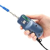
- [●400 times magnification, the fiber end face is clearly visible]
- [●Less than 1μm resolution, clear imaging.]
- [●USB Port compatible with the PCs and Laptops(Windows), OTDR( Komshine)]
- [●Applicable for UPC: SC/FC/ ST/LC/ SC Female/LC Female(Included); Applicable for APC: SC/SC Female(Optional) ]
- [●Quick focus manually. Light weight and portable, simple operation.]
What two-photon excitation added
Two-photon excitation (TPE) uses two lower-energy photons absorbed nearly simultaneously to reach an excited state that can also be reached with a single higher-energy photon. The study combined the platinum probes with near-infrared TPE, extending the imaging demonstration beyond a single excitation approach. The authors discussed deeper tissue imaging as a potential application, but the reported work does not establish a clinical imaging outcome or validate performance in human tissue.
Outdated Drivers Are Slowing You Down
One free scan finds every outdated or missing driver and matches the right update for your exact hardware.Free scan · exact hardware matchPC Slower Than It Used to Be?
A free scan shows the junk files, broken settings and background clutter dragging Windows down - then fixes them in one click.Free scan · Windows 10 & 11What the results do—and do not—say about safety and targeting
The authors described the compounds as having low cytotoxicity under the conditions they studied. That finding is limited to those experimental conditions; it does not establish broad biological safety, suitability for human use, or clinical approval. Likewise, the reported preference for nucleic-acid structures, especially nucleoli, is a localization observation in the examined cells rather than proof of a fully selective targeting system.
Rank #3
Why this was a research demonstration, not a ready-made product
The primary paper, “Time-resolved and two-photon emission imaging microscopy of live cells with inert platinum complexes,” appeared in PNAS in 2008 (doi:10.1073/pnas.0804071105). Chemistry World’s contemporary coverage described the probes as glowing for several microseconds versus a few nanoseconds for conventional probes, and noted that the dye had not then been coupled to biological molecules. Antibody conjugation and deeper imaging were presented as possible extensions, not completed clinical capabilities.
The cited work does not establish present-day commercial availability, clinical adoption, or a specific product, supplier, or microscope accessory. Its contribution is a proof of concept: long-lived emission can be used to reduce short-lived cellular background when imaging live cells.
Quick Recap
Best Value

- 1. 400 times Fiber Optic Connector Inspection Probe and 3.5 inch HD LCD display.
- 2. Fiber Optic Connector Inspection Probe Suit for FC/SC/LC/ST UPC Male Connect Head and SC/LC UPC Female Connect Head
- 3. Small size, one-handed, convenient and fast.Rotary metal bracket, stable and convenient.
- 4. 4000mAH large capacity lithium power, 20 hours standby.
- 5. Real time video,support extend t-flash card,quick check the pollution degr
Product prices and availability are accurate as of the date/time indicated and are subject to change. Any price and availability information displayed on Amazon at the time of purchase will apply.

