
Do these 3 things before closing this tab:
1Clear out junk files and repair common Windows errors2Fix the driver behind crashes, sound loss and screen glitches3Repair Windows errors before they cause bigger problemsiTechGuides is reader-supported. When you buy through links on our site, we may earn an affiliate commission. As an Amazon Associate I earn from qualifying purchases. Learn more
Yes, an MRI magnet can exert forces powerful enough to support the tank analogy—but no, it does not lift a tank during a scan. The comparison came from a 2013 description of a planned 11.75-tesla scanner. As of 1 October 2026, the strongest announced human-research MRI is the 14-tesla DYNAMIC system being built in Nijmegen, the Netherlands. It is still under construction, not a routine hospital scanner.
What does “strong enough to lift a tank” actually mean?
In 2013, IEEE Spectrum reported that the superconducting magnet planned for the INUMAC brain-imaging project would reach 11.75 teslas and described it as strong enough to lift a 60-metric-ton battle tank. That was a comparison illustrating the magnet’s force—not a claim that a scanner lifts tanks or patients during imaging. The INUMAC figures were project-era plans, not measurements of the later 14-tesla DYNAMIC system.
Tesla measures magnetic field strength. A powerful MRI magnet creates a strong static field, and magnetic materials can experience substantial forces in that field. The tank analogy conveys the scale of the magnet; it is not a practical description of how an MRI scan works.
What is the world’s strongest MRI now?
As of 1 October 2026, Radboud University describes DYNAMIC as the world’s first 14-tesla human-research MRI. Neoscan Solutions reported that magnet production began on 10 September 2026, with installation in Nijmegen scheduled for 2027. That makes it the strongest announced human-research system, not an operating scanner available for ordinary patient appointments.
#1 Best Overall

- REALISTIC DETAILS: The patient lies on the bed and listens to calming music at the PLAYMOBIL MRI with Child. The radiologist looks at the images on his monitor and makes a careful diagnosis.
- INSPIRATIONAL PLAY: Great gift for an aspiring doctor.
- COMBINE WITH OTHER HOSPITAL-THEMED SETS: (#71615-#71619 - sold separately) for more caretaking experiences.
- PLAY FUN FOR CHILDREN 4 YEARS OLD AND UP: Ideal for children's hands thanks to its age-appropriate size and safe rounded edges. Encourages your child's imagination.
- CONTENTS: Set includes one adult figure, one child figure, MRI, desk, computer, and other accessories.
The comparison below distinguishes field strength from operating status: a planned or newly manufactured magnet is not the same as a system already producing images.
| System | Field strength | Status and imaging details |
|---|---|---|
| UIC human-research MRI (2004) | 9.4 T | Unveiled for research including metabolic brain imaging; the source does not state a spatial or temporal resolution. |
| INUMAC plan (IEEE Spectrum, 2013) | 11.75 T | Planned brain imager. Project-era specifications projected 0.1 mm spatial resolution and one-tenth-second temporal resolution; these are not specifications for every current scanner. |
| CEA Iseult (CEA, 2021) | 11.7 T | Had produced first images; CEA reported 400-micron three-dimensional resolution. Its 132-ton magnet was designed for whole-body human imaging. It remains a research platform. |
| DYNAMIC, Nijmegen (Radboud University and Neoscan Solutions, 2026) | 14 T | Under construction as of 1 October 2026; magnet production began 10 September and installation is scheduled for 2027. The project has not yet reported imaging resolution. |
| Typical clinical MRI systems (Neoscan Solutions, 2026) | About 1.5–3 T | Typical clinical range; the source does not specify a single resolution or one operating status for all systems. |
The 14-tesla DYNAMIC target is higher than Iseult’s 11.7 teslas, but Iseult had already produced first images while DYNAMIC was still being built. Field strength alone does not tell you whether a system is operational, what resolution it achieves, or whether it is used for clinical care.
How does a stronger MRI help researchers see more?
An MRI scanner’s magnet aligns atomic nuclei in the body. Radio waves disturb that alignment; as the nuclei return toward their original state, they produce signals that the scanner processes into images. UIC explains that standard MRI primarily uses hydrogen signals. Its 9.4-tesla research program could also detect signals from sodium, phosphorus, carbon, nitrogen, and oxygen.
Quick wins for a faster PC:
Clear out junk files and repair common Windows errorsFree Scan →Fix the driver behind crashes, sound loss and screen glitchesFind Drivers →A stronger field can increase signal sensitivity, giving researchers a better chance of measuring subtle signals and supporting finer structural, neural-circuit, or metabolic studies. It does not mean every image automatically has finer detail: results depend on the scanner, the imaging method, and what is being measured. For example, the 0.1 mm spatial and one-tenth-second temporal figures associated with INUMAC were projections for that planned project, not a general promise about ultra-high-field MRI.
Rank #2

- EXPLORE HUMAN ANATOMY: Discover the wonders of the human body with this 8-piece set of realistic organ models, featuring replicas of the brain, heart, lungs, liver, stomach, intestines, and kidneys.
- HANDS-ON LEARNING: Engage young minds with hands-on, tactile play using these 3D anatomy figurines, fostering a deeper understanding of human biology.
- TEACHING PROPS FOR EDUCATION: Perfect for classrooms, homeschooling, or imaginative play, these anatomy toys make learning about the human body exciting and accessible for children.
- PREMIUM HAND PAINTED: Individually hand painted and crafted with attention to detail, these figurines are great for educational purposes, classroom settings, or creative play. Non-toxic and BPA free. Figurine sizes vary (1.25" to 3.75")
- PORTABLE STORAGE: Keep your parts of the body for kids organized and ready for on-the-go learning with the convenient, reusable storage tube - ideal for educators and students alike! For ages 3 and up
What might a 14-tesla scan reveal about the brain?
DYNAMIC researchers aim to map neural circuits at high resolution and study brain disorders, including changes at early stages. They also hope to examine disease metabolism and how it responds to treatment without surgery or biopsy. The project includes specialized coils and an MR dummy-scan chamber for developing and testing experiments.
These are research goals, not proof that a 14-tesla scan already diagnoses those disorders or replaces a biopsy. The broader motivation has been part of high-field MRI research for years: UIC’s 2004 9.4-tesla program proposed using metabolic imaging to study conditions including stroke, Alzheimer’s disease, autism, and mental illness, as well as responses to experimental therapies. Those are research applications, not established routine services at 14 teslas.
Independent reader supportYour contribution helps us test, update, and keep practical guides available for everyone.“With this, the world’s most powerful MRI, we can gain insight into the metabolism of diseases in humans and how these can be influenced by medication, without the need for surgery or a biopsy.”
PC Slower Than It Used to Be?
A free scan shows the junk files, broken settings and background clutter dragging Windows down - then fixes them in one click.Free scan · Windows 10 & 11Outdated Drivers Are Slowing You Down
One free scan finds every outdated or missing driver and matches the right update for your exact hardware.Free scan · exact hardware matchSpecial offer. See more information about Outbyte and uninstall instructions. Please review EULA and Privacy policy.— Dennis Klomp, professor of High-Precision Structural and Metabolic Imaging at UMC Utrecht, describing the DYNAMIC project
Rank #3
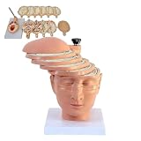 Human Brain Cross Section Anatomical Model with Digital ID, CT MRI Mould, 12 Pieces
- Precisely sculpted cross-section of the human brain, featuring 12 individually numbered pieces for clear identification of internal structures.
- Includes digital identification guide and reference to CT and MRI imaging, bridging anatomical study with clinical radiology.
- Each piece is color-coded and detachable, allowing hands-on exploration of lobes, ventricles, and major neural pathways.
- Made of durable,PVC material, easy to clean and built for repeated handling in classrooms, labs, or clinical training.
- Ideal for medical students, educators, neurologists, or as a professional gift for healthcare practitioners and anatomy enthusiasts.
Why isn’t a 14-tesla MRI a normal hospital scanner?
The projects cited here are research infrastructure. Radboud and Neoscan describe DYNAMIC as a human-research scanner under construction, while CEA describes Iseult as a research platform. Neither source establishes routine clinical use at 14 teslas. A research system can be designed to investigate what stronger fields make measurable without being an approved, widely available diagnostic service.
DYNAMIC’s design also uses high-temperature superconductors and conductive cooling. Radboud University reported €19 million in NWO Roadmap funding for the project in 2023. Those engineering choices and project funding help explain the scale of the undertaking; they do not change its stated research role or construction timeline.
How the strongest human-research MRI projects developed
- 2004 — UIC, 9.4 T: Unveiled a human-research system intended to advance metabolic brain imaging, including measurements from nuclei beyond hydrogen.
- 2013 — INUMAC, planned 11.75 T: IEEE Spectrum described the tank comparison and projected 0.1 mm spatial and one-tenth-second temporal resolution for the planned brain imager.
- 2021 — CEA Iseult, 11.7 T: CEA reported first images with 400-micron three-dimensional resolution from a system designed for whole-body human imaging.
- 2026 — DYNAMIC, 14 T: Radboud and Neoscan reported magnet production and a planned 2027 installation in Nijmegen.
So the short answer is that the tank comparison describes the forces associated with an exceptionally strong magnet, while the 14-tesla scanner points to a different promise: research measurements of brain structure, circuits, and metabolism that conventional clinical systems may not capture as sensitively. The new system’s actual imaging performance will be established after it is installed and tested.
Quick Recap
Product prices and availability are accurate as of the date/time indicated and are subject to change. Any price and availability information displayed on Amazon at the time of purchase will apply.

