
iTechGuides is reader-supported. When you buy through links on our site, we may earn an affiliate commission. As an Amazon Associate I earn from qualifying purchases. Learn more
Scientists can study activity inside a living cell with nanoscale sensors and probes, but “mini probe” is an umbrella term, not one miniature microscope. Some probes report local chemistry, some detect a chosen molecule, and others map internal structure. The right method depends on the question—and probes that enter a cell can affect it.
What does “mini probe” mean in cell research?
It describes several distinct nanoscale tools for measuring or imaging living cells. A fluorescent particle may report a chemical condition such as acidity; a functionalized fiber tip may detect a target protein; and an AFM nanoprobe may map structures inside a cell. These methods produce different kinds of evidence and are not interchangeable. A review of nanosensors for individual living cells outlines this broader range of approaches: Nanoprobes and nanobiosensors for monitoring and imaging individual living cells.
“Looking inside” can mean detecting a chemical signal, identifying a molecule, or reconstructing a physical map. It does not necessarily mean viewing the whole cell through a tiny lens.
Which methods can examine a living cell?
| Approach | What it measures or shows | How it works | Main trade-off |
|---|---|---|---|
| Fluorescent particle nanosensor | A local chemical condition, such as lysosomal pH | A dye-bearing particle enters the cell; microscopy reads its fluorescence | Requires suitable calibration and interpretation in the cell’s environment |
| AFM nanoprobe (nanoendoscopy-AFM) | Internal structure and, potentially, local mechanical properties | A narrow needle-like probe enters the cell; AFM measurements are used to build a map | Provides direct access to interior structures, but penetration and scanning can damage cells |
| Optical fiber-tip biosensor | A selected molecule or protein, demonstrated for p53 | A nanoscale tip with target-recognition chemistry detects an optical signal | Target specificity and signal interpretation matter; insertion and binding can affect measurements |
| MINFLUX fluorescence nanoscopy | Positions and distributions of labeled molecules | Uses switchable fluorophores and a donut-shaped excitation beam | Needs fluorescent labeling and specialized instrumentation; it is not a penetrating probe |
| Scanning ion conductance microscopy (SICM) | Live-cell surface topography and local properties | A fine pipette scans near the cell surface; ion-current changes help maintain distance | Can examine the surface without contact in the described approach, but does not provide the same intracellular access as a penetrating probe |
The table compares what each method is designed to measure, not a ranking of overall performance. For example, a pH sensor cannot substitute for a structural map, and a high-resolution image does not by itself identify a specific protein.
#1 Best Overall

- Integrated WF16x eyepiece:** Delivers widefield views and comfortable eye relief—no separate microscope eyepiece required.
- One-piece printed head:** Monolithic design helps maintain optical alignment and reduces shake for cleaner images.
- Aluminum alloy chassis:** Lightweight yet rigid frame improves stability when capturing photos and video.
- Drop-in 23.2 mm fit:** Inserts directly into 0.913" eyepiece tubes for a fast, tool-free setup in classrooms and labs.
- Universal spring phone clamp:** Securely holds smartphones 58–100 mm (2.28"–3.93") wide; for multi-camera phones, align the primary lens.
How do fluorescent nanosensors measure cell chemistry?
A cell can take up a fluorescent particle, which then reports its local chemical environment through changes in emitted light. A 2014 Nature Protocols method describes an approximately 60 nm polyacrylamide nanosensor designed to measure pH in lysosomes. It combines pH-sensitive fluorescein and Oregon Green with rhodamine as a pH-insensitive reference, and gives a sensor response range of pH 3.1–7.0. The reported protocol takes 2–3 weeks to carry out. See Design, calibration and application of broad-range optical nanosensors for determining intracellular pH.
Calibration is essential: fluorescence must be interpreted against known conditions before an image can be treated as a pH measurement. The method also calls for attention to fluorophore choice, the sensor’s effective measurement range, its uptake and delivery to lysosomes, and how images are evaluated. Confocal microscopy can then track lysosomal pH changes in living cells.
Rank #2

- SEE EVERY DETAIL CLEARLY: Equipped with an advanced CMOS sensor, our microscope delivers unparalleled clarity with high-definition 1600x1200 resolution. From intricate patterns to minute features, experience every detail with exceptional clarity and precision.
- PLUG AND PLAY COMPATIBILITY: Effortless compatibility with Windows 10 and Mac. Simply use the pre-installed Camera App on Windows 10 or the Photo Booth application on Mac. Additional software for both platforms is available for enhanced functionality. Contact us for further details.
- INDUSTRIAL-GRADE DESIGN: Built with solid aluminum construction, our microscope stand is of the highest quality, providing industrial-grade durability and stability. It's adjustable to meet your specific needs and built to last, ensuring reliable performance for even the most demanding tasks.
- VERSATILE USES: Ideal for hobbyists, pros, and enthusiasts alike. From coin inspection to phone repair, it handles precision tasks effortlessly. Also perfect for quality control, manufacturing, and insect examination. Seamlessly adaptable for imaging tasks, including plants, skin, and hair analysis.
- CAPTURE AND RECORD WITH EASE: Effortlessly capture live video or still images while examining your samples. The USB cable connects directly to your computer, enabling seamless data transfer and easy recording. See the small world in a whole new light!
Earlier work on PEBBLE optical nanosensors described 20 nm and 200 nm particles with fluorescent indicators trapped in a polyacrylamide matrix. That 1999 study examined delivery methods including liposomal delivery, gene-gun bombardment, and picoinjection. Those are historical research examples, not a general safety finding or a claim about a currently available product.
How does nanoendoscopy-AFM map a cell’s interior?
In nanoendoscopy-AFM, a long, narrow probe is inserted into a living cell. Atomic force microscopy measurements are then used to construct two- or three-dimensional maps of intracellular features. A 2024 study demonstrated label-free imaging of internal structures, including the nucleus and actin arrangements. Its example silicon probe was specially modified using focused ion beam milling; that probe had a base diameter below 200 nm, a tip radius below 20 nm, and a length above 10 μm. These are specifications of the demonstrated probe, not a standard product specification. See Visualizing intracellular nanostructures of living cells by nanoendoscopy-AFM.
Rank #3

- Turn any traditional microscope a digital microscope! This imager fits any microscope that accepts 23 mm or 30 mm eyepiece diameters
- 5MP CMOS SENSOR: Captures high resolution still images and 30 fps video
- USB-POWERED: Connects to your personal computer via open USB port
- SOFTWARE INCLUDED: Works with both Mac and Windows PCs
- BONUS FEATURES: New and improved software with added features like measurement, calibration, note taking and comparison (allows comparing 2 live streams)
Penetrating a cell makes viability a central part of the method. In the cited study’s viability experiments, scan areas smaller than 2 × 2 μm² did not damage cells, while areas larger than 3 × 3 μm² could disrupt and kill them. Those findings apply to that study’s experimental conditions; they are not universal safe-area thresholds. Probe fabrication, insertion, scan size, and AFM instrumentation all matter.
Can a tiny probe detect a particular protein?
Yes, in a research demonstration, an optical fiber-tip biosensor detected p53 without fluorescent labeling. A 2014 study used a tapered fiber tip below 100 nm in diameter, carrying a gold nanorod functionalized with antibodies that recognize the target. The researchers observed different p53 dynamics in single HeLa cells after ultraviolet exposure versus neocarzinostatin treatment. This was a specific experimental sensor, not evidence of a ready-to-buy consumer probe. See Nanoscale Label-free Bioprobes to Detect Intracellular Proteins in Single Living Cells.
Rank #4
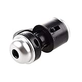
- Portable microscope works for iPhone/iPad/Samsung mobile and tablet. You can take the photo and save it on your phone.
- It fits for all smartphone or tablet thickness between 0.5inch and the distance between the camera and edge under 0.9inch.
- Built in led light, it is great for observations like coins, stamps, plants and insects.
- Magnification is 30 times.
- 3 Years Warranty Guarantee.
Recognition chemistry does not eliminate interpretation problems. The paper discusses how intracellular binding and background can affect the signal; it also reports slow p53 dissociation, which can leave residual signal after the target concentration falls. A signal therefore needs to be understood in light of the sensor’s binding behavior, not read as an instantaneous, perfectly reversible concentration measurement.
The Tool Desk
Outbyte Driver Updater FREEScan for outdated or missing drivers - takes under a minuteDriver Scan →Outbyte PC Repair FREEClear out junk files and repair common Windows errorsFree Scan →How do nanoprobes differ from nanoscopy and surface imaging?
MINFLUX is a fluorescence nanoscopy technique, not a needle or particle inserted into a cell. A 2019 Nature Methods paper reported 1–3 nm resolution for structures in fixed and living cells using switchable fluorophores and a donut-shaped excitation beam. That result belongs to the particular study and technique; it is not a routine resolution claim for all live-cell microscopes or mini probes. Fluorophore behavior, labeling, the time scale of interest, and access to specialist equipment shape whether this approach fits an experiment.
Best Value

- 100X–1000X Magnification: The 2025 upgraded digital microscope for kids supports 100X to 1000X magnification, allowing clear observation of insect wing textures, plant cell veins, intricate coin patterns, and more. It easily reveals microscopic details that are invisible to the naked eye, sparking endless curiosity about science
- 1080P HD Camera & 2.0 Inch IPS Screen: The kids microscope handheld is equipped with a 2-megapixel 1080P microscope camera, capable of capturing high-definition photos and videos. The 2.0-inch IPS screen delivers accurate colors and sharp details, presenting rich microscopic visuals in real time and enhancing the immersive observation experience. Built-in 8 LED fill lights ensure a bright and clear view even in low-light conditions
- Portable Design for Kids: Specifically designed to fit small hands, making the mini microscope comfortable and easy to hold. Buttons positioned on top allow kids to easily observe and operate at the same time. Includes a lanyard for hanging around the neck or on a backpack, ensuring effortless portability. A built-in rechargeable battery offers long-lasting power, great for both outdoor exploration and everyday learning
- Multifunctional & PC Connectable: Supports taking photos and recording videos. Built-in storage holds up to 500 photos, and memory can be expanded via a Micro SD card (not included). The pocket microscope for kids can connect to a computer for real-time viewing on a larger screen, making it easier to study, present, and share discoveries, enriching the experience of scientific exploration
- 12 Extra Slides & Perfect Gift: Includes 12 carefully selected plastic slides covering four categories: plants, vegetables, insects, and animals. Each slide showcases clear structures and beautiful forms, blending education with fun. This kids microscope toys set is both entertaining and educational, making it an ideal gift for boys and girls aged 3 to 12 on birthdays, holidays, Christmas, or as a learning reward.
SICM and related pipette methods take a different route: the pipette scans close to the cell surface without contact in the described measurement approach, using ion-current changes to maintain distance. A 2021 review discusses nanoscale imaging of dynamic cell surfaces and possible integration with local measurements or fluorescence. Unlike AFM nanoendoscopy, this surface-scanning approach does not insert a needle into the cell.
How should a researcher choose an approach?
- Define the measurement. Choose a chemical sensor for a condition such as pH, a recognition-based biosensor for a selected target, or a mapping method for physical structure. If the question concerns labeled-molecule positions at nanometer scale, fluorescence nanoscopy may be relevant instead.
- Decide whether the method must enter the cell. Particle sensors are taken up by cells; nanoendoscopy-AFM and the fiber-tip design are inserted into them. SICM, in the described approach, examines the surface without contact.
- Check how a signal becomes a result. Fluorescent sensors need calibration and localization checks; biosensors need target-specificity and background assessment; AFM and nanoscopy require reconstruction or interpretation suited to their measurement.
- Account for cell effects and equipment. Consider penetration, imaging conditions, scan area, viability checks, and the specialized fabrication or instrumentation the method requires.
These are specialized laboratory techniques, not interchangeable add-ons for a generic consumer microscope. The method should match the biological question and the expertise and equipment available.
Quick Recap
Product prices and availability are accurate as of the date/time indicated and are subject to change. Any price and availability information displayed on Amazon at the time of purchase will apply.
Quick wins for a faster PC:
Scan for outdated or missing drivers - takes under a minuteDriver Scan →Repair Windows errors before they cause bigger problemsFix Now →Fix the driver behind crashes, sound loss and screen glitchesFind Drivers →
