
Free tools Windows power users keep installed
One-click scans. No signup required.
iTechGuides is reader-supported. When you buy through links on our site, we may earn an affiliate commission. As an Amazon Associate I earn from qualifying purchases. Learn more
Do not choose whichever classifier sounds more certain, and do not treat either output as a diagnosis on its own. Ask the treating clinical team to reconcile the original MRI images, pathology material and reports, clinical history, and each tool’s intended use and limitations. The team should first confirm that both systems assessed the same lesion with appropriate inputs, then document the discordance and decide whether specialist review, additional testing, repeat sampling, or another step is appropriate.
First determine what is actually disagreeing
“Pathology classifier” and “MRI classifier” can describe several different situations. The disagreement might be between two artificial-intelligence systems, between an AI result and a radiologist or pathologist, or between an MRI interpretation and tissue diagnosis. Those are not interchangeable problems. A model may be estimating a probability, assigning a standardized imaging category, or flagging features for a professional; a pathology report may be a definitive diagnosis, a limited sample interpretation, or an indeterminate result.
Write down the exact wording, category, score, and date for each output. Do not translate labels into a shared scale unless the developers or clinical guidelines explicitly define that conversion. A category such as “probably benign,” a numerical risk estimate, and a tumor subtype are answers to different questions.
What the clinical team should do next
- Identify the clinical question. Is the MRI trying to characterize a lesion, stage known disease, find a biopsy target, or monitor a change? Is pathology intended to identify the lesion, determine its grade, establish treatment biomarkers, or rule out malignancy? A mismatch can be only apparent when the tests address different endpoints.
- Verify the patient, lesion, and dates. Confirm the patient identity, organ, side, anatomic location, accession number, and the imaging study linked to the tissue sample. Record the pathology acquisition date. A lesion can change between imaging and biopsy, especially when the interval is long, so an older scan and newer tissue result may not describe the same biological state.
- Confirm that the biopsy sampled the imaging target. Review the procedure images, localization method, needle or device position, specimen radiograph when applicable, and the pathologist’s description of the fragments. A benign result does not explain a suspicious MRI finding if the needle sampled adjacent tissue or a different focus.
- Check image and specimen adequacy. Look for motion, incomplete MRI sequences, artifacts, contrast-timing problems, low resolution, scant tissue, crush or cautery artifact, necrosis, and poor fixation. Ask whether the AI system accepts the actual sequence, scanner format, stain, slide type, or magnification used in this case.
- Review the original evidence, not just summaries. The treating radiologist, pathologist, and relevant clinician should examine the actual images and slides or digital whole-slide files, prior studies, reports, symptoms, examination findings, laboratory data, and treatment history. A score copied into a chart cannot reveal whether the model saw an artifact or whether a microscopic feature was missed.
- Obtain an appropriately staffed second review. Depending on the organ and question, this may involve a subspecialty radiologist, subspecialty pathologist, interventional specialist, surgeon, oncologist, or a multidisciplinary conference. The American College of Radiology recommends consensus review of histology, imaging, and other clinical data for discordant liver observations reported with LI-RADS categories.
- Choose any additional procedure with the treating specialists. The options may include repeat targeted sampling, additional imaging, slide recuts or stains, outside consultation, short-interval follow-up, or surgery. The choice depends on sampling accuracy, residual imaging concern, procedural risk, and the expected diagnostic yield; no single action applies to every organ.
- Document the unresolved mismatch and plan. The record should state the imaging category or impression, pathology diagnosis, pathology date, lesion location, why the findings appear discordant, who reviewed the case, what uncertainty remains, and the agreed follow-up trigger or deadline.
How to assess concordance
Concordance asks whether the tissue result plausibly explains the imaging appearance. It is not the same as asking whether either test is “right” in isolation.
#1 Best Overall
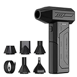
- Powerful Turbo Fan:CAGIWIRU Electric Air Duster have reached speed up to 130000RPM,Can efficient remove dust and debris.With LED light and Three Adjustable Speed to meet with different cleaning tasks.
- With Attached USB C Fasting Charging cable.Charing 3 hours,Enjoy up to 240 minutes of use on the lowest setting, For third speed can use for 25mins.with four charging options to meet with your needs.
- Wide Application: attached 5 different nozzles,making it suitable for a variety of scenes, such as car,pet, pc, keyboards, and other electronic devices. It also serves for office and home clean duster.
- Handy Design & Portable:Cordless air duster electric nozzle is Light weight and small size, design and comfortable to hold and space saving, convenient to carry around. You can put it in your room or inside the car without taking up space.
- Economical &Reusable: Built durable materials with a long-lasting battery,our CAGIWIRU air duster it's a sustainable alternative to disposable air cans.
Concordant benign result
A benign diagnosis is concordant only when the sampled tissue, lesion location, imaging morphology, and clinical setting fit together. For example, a clearly localized benign process that has the expected MRI appearance may need routine management. A generic benign label from a tiny or poorly targeted sample is not automatically explanatory.
Concordant malignant result
A malignant diagnosis may still require correlation with the MRI to establish the full extent of disease, identify additional lesions, and plan treatment. The MRI can show disease outside the sampled focus; pathology from one fragment does not classify every enhancement in an organ.
Discordant benign or indeterminate result
This is the situation that most often requires adjudication: the MRI remains suspicious, but the tissue is benign, nonspecific, or insufficient. The team should determine whether the sample was representative and whether the pathology could plausibly produce the observed imaging features. “Benign” and “no malignancy identified” are not equivalent statements when the specimen is scant or the target was uncertain.
Windows Errors? Fix Them Before They Spread
Repair common Windows errors and clear accumulated junk for a smoother, more stable PC - no reinstall needed.Free scan · no reinstallOutdated Drivers Are Slowing You Down
One free scan finds every outdated or missing driver and matches the right update for your exact hardware.Free scan · exact hardware matchRank #2

- Screen Cleaner is Your Screen’s New Bestfriend! - EVEO is proud to present a screen cleaner that’s perfectly suited for your TV. Meet your new screen cleaner, computer screen cleaner, laptop cleaner, iPad cleaner, Macbook,tv cleaner or any other electronic device. Our screen cleaner spray and wipe along with the included plush microfiber cloth, easily removes dust, fingerprints & other smudges on the screen’s surface. Clean effectively for a streak-free screen
- Gentle on your Screens - Our screen cleaner spray and wipes is compatible to be used even for the most sensitive LCD, LED, CRT, and OLED screens in proper use. Be at ease knowing that this product is compatible with all the major brands: LG, SONY, Samsung, Sharp, iPad, iPhone, Android screens, Kindle, PC monitors - we’ve got you covered! Can be used as LCD screen cleaner, tv cleaner, smart tv screen cleaner, and more
- Premium Super Soft Plush Microfiber Cloth - Included in this efficient screen cleaning kit. This screen cleaner spray and wipe has our signature plush microfiber cloth specially designed to comply with screens and monitors and leave them streak-free. Has been shown to effectively absorbs stubborn stains, zero streaks, and no fiber shedding. Always use this together with our screen cleaner for the best results
- Easy-to-Use Screen Cleaner Kit + microfiber cleaning cloth - The EVEO Screen Cleaning Kit will easily be your new favorite tool when it comes to taking care of the things you love most, quick and easy solution for a streak-free screen. Simply screw on the sprayer gun on the screen cleaner bottle. Spray the cleaning liquid onto the microfiber cloth, then wipe the screen until desired cleanliness & shine is achieved.
- Quality Screen Care Kit Worth Purchasing - Enjoy a convenient Screen Cleaner anytime, anywhere you need it. Your satisfaction is our top priority. We stand behind the quality of our products and that’s why, you shouldn’t be worrying at all. We manufacture the Best Screen Cleaner. and can be used for laptop cleaning kit, laptop screen cleaner, tv screen cleaner for smart tv. MacBook screen cleaner, monitor cleaner or screen cleaner spray for electronic devices with microfiber cleaning cloth
AI classifier checks before anyone acts on a score
The College of American Pathologists’ guidance on validating AI algorithms in anatomic pathology emphasizes intended use, output definition, and representation of relevant diagnostic entities in validation. Apply those checks to both pathology and MRI software.
| Question | Why it matters |
|---|---|
| What is the intended use? | A tool designed to flag a possible focus for review is not necessarily validated to diagnose, exclude disease, grade a tumor, or select treatment. |
| Who is the intended user? | Many systems are designed for trained professionals who independently review the source images and clinical information, not for unsupervised patient interpretation. |
| What inputs are required? | Scanner field strength, sequences, contrast phase, image format, slide preparation, stain, magnification, or tissue quality may be mandatory. |
| What population was evaluated? | Performance can vary with organ, disease prevalence, demographics, acquisition protocols, specimen preparation, and referral setting. |
| What does the label mean? | A probability, heat map, triage flag, category, and diagnostic class have different error modes and cannot be treated as interchangeable evidence. |
| Was this case inside the validated scope? | An unusual lesion, protocol, scanner, stain, or low-quality input may be outside the evidence supporting the software. |
| What information did the system not receive? | A model may not know symptoms, prior cancer, laboratory values, prior imaging, procedure details, or the pathology result itself. |
U.S. Food and Drug Administration clinical decision-support policy describes MRI and pathology images as medical images and emphasizes intended user and population, input requirements, algorithm methods, clinical validation, and patient-specific information for independent professional review. Regulatory classification or clearance does not make a particular output correct for an individual case. The exact product labeling and current version must be checked by the clinical service using it.
When sampling accuracy changes the decision
The key branch is whether the biopsy actually captured the MRI abnormality.
Rank #3

- 【Large size】:Measuring 800 x 300 mm/ 31. 5×12 inches, which is wider and taller than others. Large mouse pad can perfectly fit your keyboard and mouse of any size, and there is also ample space for peripherals such as phones, tablets, etc.
- 【LED backlight mouse pad】:Blade Hawks RGB mouse pad provides 7 static modes and 7 dynamic modes will give you the options you are looking for. Power-off memory function remembers the last lighting mode you selected. The super glow fiber Chroma will give you a colorful gaming setup that you want.
- 【Anti-slip rubber base】:Made from Natural rubber, this gaming mousepad will firmly grip your desktop, allowing you to enjoy the ultimate precision in the games you are most passionate about. It also comes with a texture that enhances this ability even further.
- 【Smooth and waterproof surface】: Micro-textured cloth surface, for extremely precise mouse control and a smooth movement. When liquid splashes on the gaming mouse pad, it forms water droplets and slides down. Will not delay your work or gameplay.
- 【Plug and play】: This mouse and keyboard pad is powered by USB, plug and play, no driver required. One button control light modes. Press one time to change the lighting mode, press twice to change the brightness, press and hold about 3 seconds turn ON/OFF.
If the target was not adequately sampled
The team may consider repeat image-guided percutaneous biopsy directed at the suspicious focus, a different approach, or another imaging method to improve localization. The risks of another procedure, the lesion’s accessibility, and the likelihood that a new sample will answer the question should be weighed explicitly.
The Tool Desk
Outbyte Driver Updater FREEScan for outdated or missing drivers - takes under a minuteDriver Scan →Outbyte PC Repair FREEClear out junk files and repair common Windows errorsFree Scan →If the target was accurately sampled but pathology does not explain the MRI
Additional tissue or a different diagnostic pathway may be necessary. In its review of breast MRI problem-solving, the Radiological Society of North America describes excisional biopsy as the standard of care for an accurately sampled lesion with radiologic-pathologic discordance, while repeat percutaneous biopsy may be attempted when sampling was inaccurate. That recommendation is specific to the breast context and should not be transferred automatically to liver, brain, prostate, or another organ.
If both tests are limited
When MRI quality and tissue adequacy are both uncertain, a specialist may first repeat image interpretation, obtain better imaging, request deeper sections or additional stains, or seek outside pathology review. A second automated score without fixing the underlying input problem rarely resolves the uncertainty.
Rank #4

- 【Softer and More Comfortable】Vaydeer wrist rest has unique diamond pattern, which is the combination of softness and aesthetics. The materials of wrist rest are improved into higher quality memory foam and covered with silky smooth lycra. The computer wrist rest makes you as comfortable and cushiony as like rest your wrists on clouds.
- 【Ergonomic Wrist Saver】The wrist rests for keyboard and mouse comes with a 17.32×3.15×0.83 inch keyboard wrist pad and a 5.94×3.15×0.83 inch mouse wrist support. Based on ergonomic design, the unique concave shape is the perfect fit for your wrist joints. The wrist rest pad fits most computer keyboards and laptops, improve hand and wrist posture, release your wrist and arm stress.
- 【Non-Slip Rubber Bottom】Featuring an anti-skid silicone base on the bottom, this wrist keyboard support stays firmly in place on your desk, preventing the padding from sliding around, ensuring stable and consistent wrist support during extended computer sessions.
- 【Better Experience & Pain Relief】Our keyboard arm rest is beneficial to alleviate the soreness caused by direct contact and friction between your arm and a hard desk surface, reducing the risk of wrist fatigue or carpal tunnel. The soft texture of memory foam can evenly distribute the pressure around your wrists and provide good support with just enough give.
- 【Helpful in Multiple Scenarios】Whether you're working, studying, writing, typing, gaming, this keyboard and mouse rest combo is an essential accessory to add comfort and support to your hands and wrists. It’s also a great gift for men, women, family, friend, coworker, gamer, teacher, etc.
Organ-specific examples and limits
Liver observations and LI-RADS
Questions such as “What if the pathology diagnosis is discordant with the LI-RADS category?” refer to the American College of Radiology’s liver-specific framework, not a universal rule for every MRI and biopsy disagreement. ACR support guidance recommends multidisciplinary discussion with consensus review of histology, imaging, and other clinical data. The report should identify the pathology acquisition date because interval lesion change can affect interpretation. LI-RADS categories should be reported with their precise definitions; they should not be treated as a pathology diagnosis.
Breast MRI
Breast management depends on the imaging feature, biopsy method, radiologic-pathologic correlation, and whether the lesion was accurately targeted. The RSNA recommendation for excisional biopsy in an accurately sampled discordant lesion is a breast-specific management statement. A breast team may instead repeat percutaneous sampling when the first procedure did not sample the imaging target.
Quick wins for a faster PC:
Scan for outdated or missing drivers - takes under a minuteDriver Scan →Repair Windows errors before they cause bigger problemsFix Now →Fix the driver behind crashes, sound loss and screen glitchesFind Drivers →Other organs
For brain, prostate, kidney, pancreas, bone, and other sites, the appropriate response varies with anatomy, lesion behavior, accessibility, patient risk, and available procedures. There is no cross-organ protocol that converts a disagreement into an automatic operation, repeat biopsy, or period of observation.
Best Value

- 【No Drill Mounted】Cable management tray can be assembled in as little as 3 minutes or less-simply. Side clips easily clampe the edge of desk, pass through the holes on the sides, and secure excess cables with baskets. Desk cable management supports inward or outward installation to meet your needs in different scenarios, and provides you with great convenience when collecting and organizing wires.
- 【Rubber Pad Protects Desk】Wire organizer under desk built-in soft rubber pads on the clamp protect your desk from scratches, so you will not damage your furniture or office.
- 【Sturdy and Beautiful】Cord management under desk is made of high-quality carbon steel and holds about 15 lbs. The wire basket has 2 holes on each side of the tray to enable your cables to pass freely. Cable baskets are suitable for desk thickness use from 0.4" to 2.4".
- 【Space Saving & Keep Organized】Desk cable management tray holds all your power cords, outlet strips, USB hubs and computer transformers hidden from your feet, and the data cables will not enter the vacuum cleaner to keep your desk clean and tidy. Desk wire organizer is also suitable for use in the kitchen and outdoors.
- 【Reduce Safety Risks】Cable tray under desk keeps all plugs away from your kids, no more worry about tripping. Using 2 holes wire tray saves extra space, helps you easily cabling and wire protection. The mesh design is not easy to accumulate dust, but avoids safety accidents caused by messy wires.
How the disagreement should be documented
A clear note prevents a score or preliminary label from being mistaken for a final clinical decision. Include:
- the exact MRI impression, sequence or study date, and lesion location;
- the pathology diagnosis, specimen description, accession and acquisition date;
- the classifier name, version, intended output, input quality, and result, when an AI tool was used;
- whether the biopsy target was confirmed and what procedure evidence supports that conclusion;
- the clinical history and prior studies that materially affect interpretation;
- the specialists who reviewed the case and whether consensus was reached;
- the reason for discordance, such as targeting error, limited tissue, interval change, artifact, or a genuinely conflicting finding;
- the next action, responsible service, timeframe, and condition that would trigger escalation.
The College of American Pathologists’ active 2026 Interpretive Diagnostic Error Reduction update states that review may be prospective or retrospective but, when undertaken to detect potential diagnostic errors, should occur before management is initiated. CAP’s broader recommendation is that anatomic pathology services develop procedures to review cases for disagreements and potential interpretive errors to improve patient care.
What patients should and should not do
- Do ask which lesion was sampled, whether the sample was considered adequate, and whether the radiologist and pathologist have explicitly called the result concordant.
- Do request clarification of the exact category, probability, or diagnosis rather than relying on a color, confidence score, or app summary.
- Do ask whether the case will receive subspecialty or multidisciplinary review and what the follow-up deadline is.
- Do not select the result that sounds more certain, assume a benign label rules out disease, or infer that a high AI score proves cancer.
- Do not arrange repeat biopsy or surgery solely from an automated output without the treating team confirming the target, indication, and expected benefit.
- Do not delay urgent care while waiting for a software re-run if symptoms or the clinical situation require prompt assessment.
What is known about disagreement rates
No reliable universal percentage describes disagreement between pathology and MRI classifiers. The rate depends on the organ, disease mix, sampling method, imaging protocol, definitions of discordance, and whether the comparison involves human interpretation or software. CAP’s interpretive-error guideline concluded that available evidence was insufficient to establish a precise prevalent rate of discrepancy or error. A statistic from another disease or modality should not be used to predict an individual patient’s risk.
Likewise, current guidance does not establish one universally best resolution method or timing rule. The defensible approach is explicit concordance assessment, verification of the target and inputs, review by the relevant specialists, and a documented plan proportionate to the clinical concern.
Quick Recap
Product prices and availability are accurate as of the date/time indicated and are subject to change. Any price and availability information displayed on Amazon at the time of purchase will apply.

