
iTechGuides is reader-supported. When you buy through links on our site, we may earn an affiliate commission. As an Amazon Associate I earn from qualifying purchases. Learn more
A 3D microfluidic liver-on-a-chip is not one standardized device. It is a family of laboratory models that places liver cells—often several human cell types—in a three-dimensional, perfused microfluidic environment. The format can reproduce selected liver functions, cell interactions, chemical exposures and mechanical conditions that ordinary two-dimensional cultures often miss, but it does not recreate a complete human liver or replace animal, clinical or regulatory evidence.
What is a liver-on-a-chip?
A liver-on-a-chip, also called a liver microphysiological system, combines living liver cells with engineered channels or chambers through which culture medium is controlled. The chip determines how nutrients, oxygen, test compounds and waste products reach the tissue, while the three-dimensional arrangement gives cells more realistic neighbors and geometry than a flat plastic well.

The term covers many designs rather than a single commercial or scientific standard. One model may use primary hepatocytes in a perfused chamber; another may combine hepatocytes with stellate, Kupffer and endothelial cells; others use spheroids, organoids, scaffolds, gradients or linked liver-and-other-organ modules. “3D” describes the tissue arrangement, while “microfluidic” describes the controlled movement of fluid around or through that tissue. Either feature can vary substantially between platforms.
Do these 3 things before closing this tab:
1Scan for outdated or missing drivers - takes under a minute2Clear out junk files and repair common Windows errors3Fix the driver behind crashes, sound loss and screen glitchesThe goal is a more informative in-vitro experiment, not a miniature transplantable organ. A chip can represent selected metabolic, toxicological or disease processes under defined conditions. Results still depend on the cell source, architecture, media, flow regime, exposure protocol, assay readout and validation for the particular question.
#1 Best Overall

- ◓DESIGN: Cross-junction microfluidic chip designed for precise droplet generation and microsphere formation in laboratory settings
- ◓There are design drawings in the picture, please check and compare carefully before buying to ensure that the chip design meets your needs
- ◓APPLICATION: Perfect for fluid dynamics research, particle synthesis, and microfluidic experiments in research laboratories
- ◓CONFIGURATION: Features integrated channels in a cross-pattern layout for controlled fluid mixing and droplet formation
- ◓DIMENSIONS: Compact size suitable for standard microscope stages, with microchannels precisely engineered for consistent results
How the model is built
Liver cells and supporting cell types
Hepatocytes perform many of the liver’s central metabolic and synthetic functions, but hepatocytes alone do not reproduce the tissue environment. Researchers may add non-parenchymal cells, including stellate cells, Kupffer cells and endothelial cells, to model inflammation, extracellular-matrix interactions, immune signaling and the interface with blood vessels.
NIH NCATS describes a University of Pittsburgh model in which hepatocytes, stellate cells, Kupffer cells and endothelial cells self-assemble into plate-like cords. The arrangement is intended to create cell-to-cell relationships that are difficult to maintain in a conventional monolayer. The exact ratio, source and maturation state of each cell type are design choices, not universal requirements.
Cell sources can include primary human cells, donor-derived cells, stem-cell-derived hepatocyte-like cells or combinations of these. Each source brings trade-offs in availability, donor variability, maturity, genetic background and reproducibility. A result from one source should not automatically be generalized to every human population or disease state.
PC Slower Than It Used to Be?
A free scan shows the junk files, broken settings and background clutter dragging Windows down - then fixes them in one click.Free scan · Windows 10 & 11Crashes, No Sound, or Screen Glitches?
Random freezes, missing sound and display glitches usually trace back to one bad driver. Find and replace yours safely.Free scan · under a minuteMicrochannels, chambers and perfused medium
Microfabricated channels or chambers deliver culture medium and test compounds while removing waste. Perfusion can establish concentration and oxygen gradients, support longer exposures and impose fluid-related mechanical cues. The flow driver, channel dimensions, residence time and shear conditions differ by platform and should be reported with the experiment.
Some designs place cells directly in a channel; others keep a 3D tissue compartment beside a perfusion channel and rely on diffusion across a porous barrier or matrix. Linked modules can connect a liver compartment to another tissue so researchers can examine how hepatic metabolism changes an effect elsewhere in the model. A connected system is still an engineered experiment, not proof of what will happen in a patient.
Rank #2
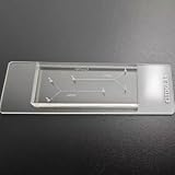
- VERSATILE STANDARD CHIP: Dual-width channels (50µm & 100µm) in a simple layout — perfect for proof-of-concept experiments and protocol development.
- ENTRY-LEVEL & AFFORDABLE: The most cost-effective way to start microfluidics — ideal for students, teaching labs and teams testing new protocols.
- STARTER KIT INCLUDED: Comes complete with PTFE tubing and a syringe, giving students and researchers everything needed to begin microfluidics experiments right out of the box.
- QUALITY PDMS/GLASS BUILD: RTV615 PDMS plasma-bonded to borosilicate glass; optical clarity for fluorescence and brightfield imaging.
- PRACTICAL FORM FACTOR: Compact 45x20mm size with multiple inlet/outlet ports for flexible fluid routing.
3D organization and biosensors
Researchers can organize cells as self-assembled cords, spheroids, organoids or scaffold-supported tissues. Biochemical gradients and matrix composition influence oxygen availability, polarity and differentiation. A design that works for a short toxicity pulse may not be suitable for a month-long disease model.
The NCATS-described model also uses fluorescent biosensor cells. Their signals can indicate events such as cell death or damage associated with free radicals, adding a dynamic warning signal to conventional measurements of viability, enzymes or gene expression. Biosensors are readouts, not a guarantee that every relevant injury mechanism is captured.
The Tool Desk
Outbyte Driver Updater FREEFix the driver behind crashes, sound loss and screen glitchesFind Drivers →Outbyte PC Repair FREEClear out junk files and repair common Windows errorsFree Scan →Materials and fabrication
Polydimethylsiloxane (PDMS) has been widely used because it is transparent, gas-permeable and convenient for rapid prototyping. Those advantages help with microscopy and early design iteration. PDMS can also absorb some small molecules, and its fabrication and bonding steps can complicate scale-up. The extent and practical importance of adsorption depend on the compound, material formulation, channel geometry and assay conditions; a generic PDMS chip should not be assumed to be assay-ready.
Other approaches include thermoplastics, 3D printing, digital light processing and bioprinting. These methods can support more customized architectures or manufacturing workflows, but they introduce their own questions about optical access, residual materials, surface chemistry, sterility and reproducibility. Material selection belongs in assay validation, especially when a hydrophobic or highly potent test compound is involved.
How a 3D microfluidic liver model works
- Define the biological question. Decide whether the experiment concerns metabolism, clearance, chronic disease, immune interaction, acute toxicity, drug-induced liver injury (DILI) or another endpoint. The question determines the required cell types, exposure duration and readouts.
- Select and characterize the cells. Document donor or cell-line origin, passage or differentiation state, expected liver functions and the presence of supporting cells. Confirm identity and baseline viability before loading the device.
- Choose the architecture. Select a cord, spheroid, organoid, scaffold or other 3D format, then specify where each cell population is located. Record matrix composition, compartment dimensions and any membrane or barrier between tissue and flow.
- Establish perfusion. Connect the chip to its medium circuit and verify that the intended flow reaches every compartment. Measure or calculate the flow conditions used in the experiment rather than describing the device only as “dynamic.”
- Allow stabilization. Give the tissue time to attach, self-assemble or reach a defined functional baseline. The appropriate period varies by cell source and design; it must be demonstrated with the model’s own viability and function measurements.
- Introduce the test condition. Add a drug, metabolite, food chemical, inflammatory signal or disease-related perturbation at a documented concentration and exposure schedule. Include vehicle, untreated and, when appropriate, positive-control conditions.
- Collect multiple readouts. Combine imaging with measurements such as secreted enzymes, albumin or urea production, gene expression, metabolic conversion, barrier integrity and cell-death signals. A single fluorescent or viability endpoint rarely explains mechanism by itself.
- Interpret against controls and validation data. Separate effects caused by the test compound from losses caused by bubbles, channel blockage, material adsorption, contamination or declining tissue function. Compare the model with an appropriate reference system before making claims about predictive value.
What researchers use liver-on-a-chip models for
Liver disease modeling
Three-dimensional co-cultures can examine interactions among hepatocytes, stellate cells, Kupffer cells and endothelial cells during inflammation, fibrosis-like remodeling or other disease processes. The controlled environment allows researchers to vary one signal at a time and observe how cell communication changes over a defined period. Disease models remain models of selected mechanisms; they do not reproduce every systemic factor present in a person.
Rank #3
- 【Microfluidic Chip Packaging Film】 Full transparency, low temperature flexibility, good air permeability, thermal and chemical stability
- 【Microfluidic Chip Packaging Film】 Shore Hardness (A): 50, tensile strength (MPa): 4, tear strength (KN/m): 7
- 【Microfluidic Chip Packaging Film】 Elongation at break (%): 100, Elastic modulus: 1.7, Refractive index: 1.41, Transmittance (%): >95(2mm)
- 【Microfluidic Chip Packaging Film】 Using temperature range (C): -40 to 200, Dielectric strength (kV/mm): 12, Dielectric constant (1MHz): 2.7
- 【Microfluidic Chip Packaging Film】 1 Pcs/package
Drug metabolism and clearance
Perfused liver tissue can be exposed to a parent compound and monitored for disappearance, metabolite formation or changes in liver-specific function. Longer or repeated dosing schedules may reveal effects that a short static culture misses. Interpretation requires accounting for dosing concentration, protein binding, medium composition, flow rate and the capacity of the cells to maintain metabolic activity.
Free tools Windows power users keep installed
One-click scans. No signup required.
Drug-induced liver injury and hepatotoxicity
DILI studies look for injury caused directly by a compound or by metabolites generated during hepatic processing. A multi-cell model can add inflammatory and structural context to hepatocyte measurements. Researchers may compare compounds within the same pharmacological class, examine dose and time dependence, and test whether a candidate produces stress signals before widespread cell death.
A chip result is not a clinical prediction by itself. The strength of a DILI claim depends on the compound panel, controls, reproducibility, biological endpoint and evidence that the model performs in the intended decision context.
Drug screening and lead selection
Chips can be used to rank candidates, investigate mechanisms and identify compounds that warrant further testing. Throughput varies widely: a highly instrumented single-chip experiment may provide richer imaging, while a plate-compatible format may support more parallel conditions. Screening capacity should therefore be judged together with data quality and the assay’s ability to maintain liver-specific function.
Food-chemical research and linked-organ studies
The U.S. Food and Drug Administration describes work evaluating organ-chip technology to better understand how food chemicals affect the human body. Other linked systems connect liver processing with another tissue compartment to study downstream effects. These applications can reveal relationships that isolated liver cells cannot, but the result remains evidence from a defined model system rather than a direct measurement of human exposure.
Can a liver-on-a-chip predict DILI?
It may improve preclinical DILI assessment in a validated use case, but no liver chip should be presented as a universal predictor. On September 24, 2024, FDA announced that its Center for Drug Evaluation and Research accepted the first letter of intent into the ISTAND pilot program for an organ-on-a-chip technology intended to study DILI for certain drug candidates. FDA said the human Liver-Chip uses four human liver cell types and compares relative DILI risk with a drug from the same class.
Acceptance of a letter of intent was the first step in a three-step qualification process. It was not completed qualification, general FDA approval, or blanket acceptance of liver-chip data for every drug program. Any regulatory use depends on the specific technology, intended context of use, performance evidence and subsequent review.
Jeffrey Siegel, M.D., director of CDER’s Office of Drug Evaluation Science, said: “DILI is a leading reason drugs do not progress through the IND process, and emerging technologies like MPS show promise in assessing risks of hepatotoxicity in preclinical phases of drug development.”
How to compare liver-on-a-chip platforms
The 2025 review in Frontiers in Lab on a Chip Technologies names Emulate Liver-Chip, CN Bio PhysioMimix and Mimetas OrganoPlate among commercial examples. It discusses differences in cell composition, perfusion arrangements, imaging and throughput, but it is not a current product catalog. Confirm specifications, availability, support and validation directly with the manufacturer before committing to a study.
Recommended Free Tools
| Selection question | Why it matters | Evidence to request |
|---|---|---|
| Which cells and cell sources are included? | Cell composition affects metabolism, immune signaling, fibrosis-related responses and donor variability. | Source, donor information, passage or differentiation state, and characterization data. |
| Is the tissue perfused? | Flow changes delivery, gradients, waste removal and mechanical cues. | Flow-driving method, operating range, channel layout and proof that all compartments receive medium. |
| What duration is supported? | A short exposure assay and a chronic disease experiment have different stability requirements. | Demonstrated functional stability for the intended exposure schedule, not a generic duration claim. |
| Which readouts are available? | Imaging, secreted proteins, metabolic conversion and injury signals answer different questions. | Validated assay protocols, sampling access, sensor compatibility and controls. |
| Does the format fit the workflow? | Throughput, microscopy access and automation determine practical study size. | Number of conditions per run, plate or instrument compatibility, and liquid-handling requirements. |
| What validation supports the intended use? | A platform validated for metabolism may not be validated for chronic inflammation or DILI. | Performance data, reference compounds, reproducibility statistics and a clearly defined context of use. |
Limitations and common failure modes
Loss of long-term liver function
Maintaining mature, liver-specific function for extended periods remains difficult. Cells can dedifferentiate, proliferate unevenly or respond to the matrix and medium in ways that alter metabolism. Long experiments need scheduled checks of function, not only a viability measurement at the end.
Best Value
- 【Microfluidic Chip Packaging Film】 Full transparency, low temperature flexibility, good air permeability, thermal and chemical stability
- 【Microfluidic Chip Packaging Film】 Shore Hardness (A): 50, tensile strength (MPa): 4, tear strength (KN/m): 7
- 【Microfluidic Chip Packaging Film】 Elongation at break (%): 100, Elastic modulus: 1.7, Refractive index: 1.41, Transmittance (%): >95(2mm)
- 【Microfluidic Chip Packaging Film】 Using temperature range (C): -40 to 200, Dielectric strength (kV/mm): 12, Dielectric constant (1MHz): 2.7
- 【Microfluidic Chip Packaging Film】 1 Pcs/package
Perfusion, bubbles and channel occlusion
Stable flow is essential but fragile. Air bubbles can interrupt delivery and change the exposed surface area; debris or cell overgrowth can narrow channels. A run that experiences an undocumented flow interruption may not be comparable with a properly perfused run. Record pressure or flow observations, inspect channels and define exclusion criteria before analyzing results.
Standardization and reproducibility
Different laboratories may use different cell sources, matrices, media, channel dimensions and dosing schedules while calling all of them “liver-on-a-chip.” Those differences can change the outcome. Protocols should specify every variable that affects transport or biology, and reference compounds should be included when comparing runs or sites.
Scale and throughput
More biological complexity often means more handling steps, imaging and quality-control work. A platform that provides detailed single-tissue measurements may not deliver the number of conditions needed for a large screen. Conversely, a high-throughput format may offer fewer cell types or less spatial information. Throughput is a design trade-off, not a standalone quality score.
Biological and material confounders
Donor variability, immature stem-cell-derived hepatocytes, adsorption to chip materials and differences in oxygenation can all influence apparent drug effects. Include material-only and cell-free controls when adsorption or nonspecific loss is plausible. Do not assume that findings from one PDMS formulation or one donor apply to every chip or patient.
What a careful conclusion looks like
A defensible report states which liver functions were demonstrated, for how long, under what flow and exposure conditions, and with which cell sources. It identifies the readouts that changed and separates direct evidence from interpretation. The conclusion should name the model’s intended context—such as metabolism screening, a disease mechanism or a DILI comparison—rather than claiming that the chip predicts all human liver responses.
For readers choosing a system, the practical decision is to match biological complexity and measurement quality to the question. Start with cell composition, perfusion, assay duration, readouts, workflow compatibility and validation. Treat the device as a complementary preclinical model whose value comes from a well-defined experiment, not from the word “3D” or “microfluidic” alone.
Quick Recap
Product prices and availability are accurate as of the date/time indicated and are subject to change. Any price and availability information displayed on Amazon at the time of purchase will apply.
What’s actually slowing this PC down?
Pick the symptom - the matching free tool is one click away.

