
iTechGuides is reader-supported. When you buy through links on our site, we may earn an affiliate commission. As an Amazon Associate I earn from qualifying purchases. Learn more
Designed protein origami can fold and assemble into precise three-dimensional cages with stability and folding kinetics similar to natural proteins, but it does not recreate a natural enzyme’s catalytic function. In a 2017 Nature Biotechnology study, Ljubetič and colleagues built more than 20 single-chain cages from modular coiled-coil protein dimers. The cages formed tetrahedra, four-sided pyramids and triangular prisms; the largest exceeded 700 amino-acid residues and measured 11 nanometres across. The same work demonstrated tetrahedral assembly in bacteria, mammalian cells and mice without evidence of inflammation in those demonstrations.
The phrase “synthetic origami” is therefore best understood as a design strategy: researchers encode a target molecular architecture into protein building blocks so those blocks self-assemble. The comparison with natural proteins concerns folding behaviour, stability and resulting shape. It should not be read as proof that the cages are enzymes, that they fold by exactly the same route as biological proteins, or that they perform natural catalytic chemistry.
What synthetic origami means in protein engineering
Protein origami is the deliberate construction of a three-dimensional protein object from parts whose interactions have been designed in advance. Instead of starting with a natural protein and asking what shape its amino-acid sequence happens to produce, a designer specifies a geometry and then chooses sequences that should assemble into it.
Free tools Windows power users keep installed
One-click scans. No signup required.
The “origami” analogy refers to programmed shape formation, not to paper-like material or to a single universal folding algorithm. A protein chain still obeys chemistry: hydrophobic interactions, electrostatic forces, hydrogen bonding, steric constraints and solvent effects determine whether the intended structure forms. Design software and laboratory measurements narrow the possibilities, while the molecules themselves carry out the final assembly.
#1 Best Overall
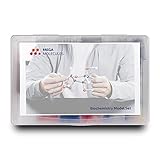
- Perfect for Visual Learners - This hands-on Biochemistry Model Kit is a tool that doesn’t just show you molecular structures; it lets you explore the magic of bonding, resonance, and chemical interactions like never before. It’s your key to understanding how molecular 3D structures influence chemical properties, such as in nucleotides and their base pairs, or in lipids and their hydrophobic behavior. You'll connect the dots between a compound's physical and chemical properties and its three-dimensional structure, deepening your understanding and making complex concepts intuitive.
- Versatile & Suitable for All Learning Levels - Whether you're a biochemistry student, an educator, or just passionate about molecular science, this model set will elevate your learning experience. It’s a powerful way to transform abstract molecular diagrams into tangible, interactive models you can touch, build, and explore. Get ready to turn curiosity into mastery—because biochemistry isn't just a subject, it's the science of life itself. With the Biochemistry Model Set, the possibilities are endless. Don’t just learn it—build it, see it, and truly understand it!
- High-Quality, Durable Components - Components are color-coded to international standards, with scaled bond lengths for accuracy. Rigid bonds allow easy single-bond rotation, while flexible bonds are ideal for double and triple bonds. Lost parts? No problem—Mega Molecules offers replacement atoms and bonds to keep your kit complete and long-lasting.
- Easy Assembly & Disassembly - Unlike many competitor kits that require special tools and make loud popping sounds during assembly or disassembly, Mega Molecules Model Sets offer a quiet, hassle-free experience. Atoms and bonds connect with a gentle push-and-twist motion and easily disconnect with a pull-and-twist—no noise, no tools needed. This thoughtful design minimizes distractions, making it perfect for classrooms, study sessions, and testing environments, ensuring a smooth, focused learning experience.
- Satisfaction Guarantee - Backed by a refund or replacement policy, this biochemistry building set offers a risk-free investment in chemistry education. Whether you're studying the protein structure, the base pairing in DNA, or the intricacies of a glycosidic linkage, this set brings your biochemistry lessons to life. Use it to model the relationships between carbon, hydrogen, oxygen, nitrogen, phosphorus, and sulfur as they form covalent bonds. With every build, you’ll gain deeper insights into the connections between molecular structure and function.
Why coiled-coils are useful modules
Ljubetič’s system used protein coiled-coil dimers as repeatable building blocks. A coiled-coil is a helical interaction module in which two or more alpha helices wrap around one another. By selecting which helices pair and by controlling the angles between connected modules, the researchers could treat each dimer as a defined edge or strut in a larger object.
Those modules were connected in single polypeptide chains. When the chains were produced, the programmed pairings brought distant portions together and closed the cage. The approach resembles assembling a geometric framework from parts with compatible connectors: the connectors provide specificity, while the overall topology determines whether the result is a tetrahedron, pyramid or prism.
What the 2017 cage study actually demonstrated
The primary report is a specific demonstration, not a field-wide size or performance limit. Its semi-automated platform and coiled-coil toolbox produced and characterized more than 20 single-chain cages in three polyhedral forms.
| Result | Reported value or observation | What the figure applies to |
|---|---|---|
| Number of characterized cages | More than 20 | The cage set in Ljubetič and colleagues’ 2017 study |
| Polyhedral forms | Tetrahedra, four-sided pyramids and triangular prisms | The same designed protein-cage platform |
| Largest construct | More than 700 amino-acid residues | The largest cage reported in that study |
| Largest measured dimension | 11 nm in diameter | The largest cage reported in that study |
| Cellular and animal demonstrations | Tetrahedral assembly in bacteria, mammalian cells and mice | Demonstrations described in the 2017 paper |
| Inflammation observation | No evidence of inflammation in those demonstrations | Those reported experimental demonstrations, not a general safety conclusion |
The paper’s authors summarized the physical comparison this way: “Their stability and folding kinetics were similar to those of natural proteins.” That sentence describes the designed cages tested in that study. It does not establish that every synthetic protein origami design behaves like a natural protein, nor that the cages have natural proteins’ biological roles.
Rank #2
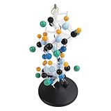
- [VIBRANT COLORS & SAFE DESIGN] The protein molecular model features bright and standardized atomic colors making it visually appealing and easy to identify. The high quality plastic construction ensures durability and safety for classroom use. The colorful balls and connecting are well made providing a tactile learning experience for students.
- [3D MOLECULAR VISUALIZATION] This model offers a comprehensive 3D representation of protein structures allowing students to study molecular and angles from all directions. The detailed design helps in understanding complex protein configurations making it an excellent tool for biochemistry and biology education.
- [HANDS ON LEARNING TOOL] Perfect for building both common and slightly complex protein structures this kit enhances interactive learning in classrooms and laboratories. The easy to assemble and disassemble design with included disconnect tool encourages student participation and engagement.
- [PORTABLE & USER FRIENDLY] Designed for convenience this molecular model is lightweight and easy to store or transport. The tightly held atoms and ensure stability during use while still allowing for effortless disassembly making it ideal for repeated classroom demonstrations.
- [EDUCATIONAL ENHANCEMENT] Specifically tailored for the QH3212 2 protein molecule this model aligns with textbook structures to boost student interest and comprehension. Teachers can leverage this visual aid to deliver more intuitive and effective lessons in molecular biology.
How a designed cage folds and assembles
A natural protein usually begins as a linear amino-acid chain whose sequence was shaped by evolution. As it emerges from a ribosome, local segments form secondary structures and distant parts find a lower-energy arrangement. The sequence, cellular environment and available helpers influence the route and the final state.
In the designed system, the sequence is selected to favour a predetermined network of contacts. The chain contains coiled-coil segments that preferentially pair with their intended partners. Those interactions can draw separate regions together, close a polyhedral face and ultimately produce a cage. The design goal is not merely “a stable protein,” but a stable protein with a chosen connectivity and cavity.
Folding versus assembly
For a single-chain cage, folding and assembly are intertwined. Individual helices must form, the correct coiled-coil partners must meet, and the complete chain must avoid kinetically trapped alternatives. A cage can therefore be thermodynamically favourable yet difficult to reach if an early mispair blocks a later connection.
Recommended Free Tools
This is where the natural-protein comparison needs care. Similar folding kinetics mean that the measured rates and stability of the reported constructs were in the range expected for natural proteins under the study’s conditions. It does not mean that the energy landscapes, cellular quality-control pathways or evolutionary histories are identical.
Rank #3

- FOR BASIC TEACHING TO ADVANCED SCIENCE: 444 pieces molecular model kit, including 136 atoms, 158 bonds and 150 parts for Carbon-60(Fullerene), provides to students from Grade 7 to Graduate level.
- TWO CHEMICAL STRUCTURE MODELS: The ball-and-stick models use spheres to represent atoms and sticks to represent chemical bonds. In the space-filling model, the spheres are drawn to scale and are next to one another as atoms are in real molecules.
- CHEMISTRY EDUCATIONAL MOLECULE MODEL IN 3D: It can display chemical structure, molecular bond, and bond angle in all directions. Demonstrate fundamental molecular geometry, chemical molecular structure, stereochemistry with 3D modeling studies.
- EASY TO LEARN: The universal standard adopted for each atom's color makes it easier for you to use and learn. Atoms and chemical bonds combine tightly and firmly and can be easily disassembled by disconnecting tools.
- If you’re not in love with it for whatever reason, we’ll give you a full replacement or refund—no questions asked. If you have any doubt, please tell us. With nothing to worry about, or even to share with your friends, try it now.
Can designed molecules fold into structures like natural proteins?
Yes, in the limited structural and physical sense demonstrated by the cage study. Designed chains folded into reproducible, nanoscale architectures, remained sufficiently stable for characterization and assembled in biological settings. The resulting cages were protein objects rather than static computer models.
No, if “like natural proteins” means biologically equivalent. Natural enzymes are specialized proteins whose three-dimensional folds position catalytic residues, bind substrates and stabilize transition states. The reported cages were designed as containers and geometric frameworks. The study did not show that they catalyse reactions, reproduce an enzyme active site or carry out a metabolic pathway.
What the analogy captures
- Both systems use amino-acid sequences whose interactions drive spontaneous folding.
- Both can occupy defined three-dimensional structures rather than random coils.
- Both have folding rates and stability that can be measured experimentally.
- Both may encounter misfolded or kinetically trapped states before reaching the preferred structure.
Where the analogy stops
- Natural proteins were shaped by biological evolution; synthetic cages are engineered for a specified geometry.
- A cage’s structural stability is not evidence of catalytic activity.
- Natural proteins can undergo regulated conformational changes, bind many partners and participate in pathways; those functions were not established for the reported cages.
- Results from one set of coiled-coil designs cannot be generalized to every protein-origami architecture.
How researchers design a protein-origami object
- Choose the target geometry. The designer starts with a polyhedron or another architecture and defines its vertices, edges, faces, symmetry and approximate dimensions.
- Select compatible modules. Coiled-coil dimers or other interaction units are chosen so that each intended connection is strong and specific while unwanted pairings are minimized.
- Encode the connectivity. The modules are arranged in a single-chain sequence or another defined construct. The order must allow the chain to reach every required contact without excessive strain.
- Model the structure. A semi-automated design workflow checks whether bond angles, interfaces and chain routing are geometrically plausible. Computational predictions are hypotheses, not substitutes for experiments.
- Produce the protein. Constructs are expressed and purified, then examined for aggregation, degradation and correct molecular mass.
- Verify shape and assembly. Biophysical and structural measurements determine whether the intended cage forms, how uniform the particles are and whether the dimensions match the design.
- Measure stability and kinetics. Researchers test how the cage responds to temperature, chemical conditions and time, and how quickly it reaches its folded state. These are the measurements behind the comparison with natural proteins.
- Test the intended setting. If biological use is contemplated, the construct must be examined in cells or animals rather than assumed to behave the same way in a purified test tube.
Why folding pathways matter
A successful design must control not only the final shape but also the route to that shape. A molecule that can eventually reach the target may still fail in practice if it forms stable wrong contacts first. Cooperative assembly can help: once several correct contacts form, they can make the remaining contacts more favourable.
What’s actually slowing this PC down?
Pick the symptom - the matching free tool is one click away.
Work on DNA-origami folding pathways offers a useful design analogy. A DNA nanostructure study found that assembly was cooperative, that reversible bond formation helped structures recover from transient misfolding, and that forming some long-range connections early could guide particular folds. Those findings concern nucleic-acid nanostructures, not the protein cages in the 2017 report. They illustrate a general principle: reversible, staged and strategically ordered interactions can improve folding reliability.
Rank #4
- Build common and slightly complex protein structures with this molecular model kit, perfect for teaching and lab use.
- Brightly colored balls and connectors follow standard color coding, making it easy to identify and use.
- Ideal for demonstrating protein molecular features from textbooks, boosting student interest and helping teachers explain clearly.
- 3D modeling shows basic molecular structure from all angles, helping visualize molecular and angles.
- Easy to learn, store, and carry; atoms and connectors fit tightly yet can be easily disassembled with a disconnect tool.
DNA origami is related, but chemically different
DNA origami programs shape with nucleic-acid strands, commonly using a scaffold strand and many shorter staple strands. Protein origami uses polypeptide chains and engineered protein interfaces such as coiled-coils. Both approaches turn molecular sequence information into spatial organization, but their chemistry, flexibility, error modes and production methods differ.
| Comparison axis | Protein origami | DNA origami |
|---|---|---|
| Primary building blocks | Polypeptide chains, including designed coiled-coil dimers | Nucleic-acid scaffold and staple strands |
| Typical design objective in the cited examples | Self-assembled polyhedral protein cages | Defined nanocompartments that organize molecular reactions |
| Assembly mechanism | Programmed protein–protein contacts and chain folding | Complementary base pairing and scaffold–staple hybridization |
| Demonstrated setting | Purified constructs plus bacterial, mammalian-cell and mouse demonstrations | Cell-free enzyme-reaction model |
| Function shown in the cited work | Structural assembly and cage stability | Spatial confinement and coupling of enzyme reactions |
Calling both methods “origami” highlights programmable architecture; it does not make their materials interchangeable. A DNA cage cannot be treated as a protein cage with a different label, and performance in one platform cannot be transferred automatically to the other.
Independent reader supportYour contribution helps us test, update, and keep practical guides available for everyone.What the 2024 DNA-origami enzyme study adds
A 2024 Nature Nanotechnology study illustrates a different use of programmable molecular shape. Researchers connected two DNA-origami nanocompartments, each measuring 25 × 41 × 53 nanometres. One compartment housed p97, a protein-unfolding machine; the other housed α-chymotrypsin, a protease.
PC Slower Than It Used to Be?
A free scan shows the junk files, broken settings and background clutter dragging Windows down - then fixes them in one click.Free scan · Windows 10 & 11Outdated Drivers Are Slowing You Down
One free scan finds every outdated or missing driver and matches the right update for your exact hardware.Free scan · exact hardware matchIn the cell-free model, confinement increased individual reaction rates by up to tenfold. Connecting the compartments coupled the sequential steps and reduced off-target proteolysis by almost sixfold. The architecture acted as a synthetic nanofactory: it brought selected components together while limiting unwanted exposure elsewhere.
Best Value

- Model the molecular mechanics of gene expression — from DNA to protein. The Protein Synthesis Molecular Model Set from Mega Molecules is a hands-on educational tool designed to guide students through the complete process of protein synthesis: transcription and translation. Using color-coded components, this set allows learners to construct and manipulate accurate physical models of DNA, mRNA, tRNA, and amino acids—making the molecular biology behind gene expression tangible and engaging.
- This model set supports an active learning experience in which students construct DNA nucleotides using phosphoric acid, deoxyribose, and the four nitrogenous bases: adenine, thymine, cytosine, and guanine.
- Users build a DNA strand from a gene sequence (e.g., T-A-C-C-T-G-C-A-G-A-C-T), physically connecting the nucleotides via gray bonding links to represent covalent bonds.
- Users transcribe mRNA by pairing RNA nucleotides (adenine, uracil, cytosine, guanine) to the DNA template, demonstrating base pairing rules (e.g., A–U, C–G).
- Users model tRNA molecules with built-in anticodons and specific amino acid attachments—highlighting how tRNA ensures accurate translation at the ribosome.
Those results do not show that the protein cages from the 2017 study are enzymes. They show that DNA origami can organize enzymes and influence pathway behaviour through proximity and compartmentalization. The two studies address complementary questions—how to build a protein cage and how to arrange enzymes in DNA compartments—rather than testing the same performance measure.
What these findings could enable
Defined nanoscale containers
Protein cages may provide genetically encodable containers whose dimensions and interfaces can be tuned through sequence design. A cage could, in principle, present binding sites, shield a payload or organize several components. Each proposed use would require separate evidence for loading, release, stability, distribution and biological compatibility.
Design rules for self-assembly
The coiled-coil toolbox offers a way to study how modular interactions produce predictable geometry. Because the building blocks are proteins, researchers can express them biologically and alter their sequences rather than manufacturing every component by chemical synthesis.
Bridging structure and function
The long-term challenge is to add function without losing the folding reliability of the scaffold. A functional cage might need catalytic residues, recognition sites or responsive hinges, all of which introduce new interactions and possible failure modes. Structural success is the starting point for that work, not proof that a useful biological device already exists.
Important limits and how to interpret the evidence
- One principal protein study: The headline numbers—more than 20 cages, three shapes, over 700 residues and 11 nm—belong to the 2017 Ljubetič study. They are not universal limits for protein origami.
- Different applications: The 2024 DNA-origami result concerns enzyme reactions in cell-free compartments, not catalytic activity by protein-origami cages.
- Laboratory-stage evidence: The demonstrations are experimental nanotechnology research, not an approved treatment, consumer product or established manufacturing process.
- Safety remains context-dependent: No inflammation was observed in the reported bacterial, mammalian-cell and mouse demonstrations, but that observation does not establish safety for every sequence, dose, cargo or clinical route.
- Analogy is not identity: Similar stability or folding kinetics do not prove that a synthetic construct has the same mechanism, dynamics or biological role as a natural protein.
Bottom line for readers
Synthetic protein origami has crossed the boundary from conceptual drawings to experimentally assembled molecular objects. The 2017 work showed that carefully designed coiled-coil modules can produce nanoscale polyhedral cages and that those cages can display stability and folding kinetics comparable to natural proteins under the tested conditions. That is a meaningful structural achievement.
It is not evidence that the cages are natural enzymes or that engineered folds reproduce biological catalysis. DNA origami supplies a related but distinct route to molecular organization, demonstrated in 2024 by compartmentalizing p97 and α-chymotrypsin to accelerate selected reactions and suppress off-target proteolysis. Together, the studies show how sequence-programmed matter can control shape and proximity while leaving the harder question—reliable, useful function in realistic biological environments—for further experiments.
Quick Recap
Product prices and availability are accurate as of the date/time indicated and are subject to change. Any price and availability information displayed on Amazon at the time of purchase will apply.
Do these 3 things before closing this tab:
1Clear out junk files and repair common Windows errors2Scan for outdated or missing drivers - takes under a minute3Repair Windows errors before they cause bigger problems
