
The Tool Desk
Outbyte Driver Updater FREEFix the driver behind crashes, sound loss and screen glitchesFind Drivers →Outbyte PC Repair FREEClear out junk files and repair common Windows errorsFree Scan →iTechGuides is reader-supported. When you buy through links on our site, we may earn an affiliate commission. As an Amazon Associate I earn from qualifying purchases. Learn more
PSMA PET is the molecular part of the examination. It maps where an injected, radiolabeled ligand binds to prostate-specific membrane antigen (PSMA). PET/CT adds X-ray anatomy, while PET/MRI adds MRI anatomy. These names therefore describe different combinations of the same molecular-imaging concept, not three automatically different radiotracers or interchangeable tests.
For many established prostate-cancer decisions, professional guidance addresses PSMA PET/CT. PET/MRI is available or reported in some settings, but the joint EANM/SNMMI procedure guideline reviewed here specifically covers PET/CT and says PET/MRI protocols are outside its scope. The appropriate examination depends on the clinical question, the required anatomic detail, local expertise and availability, and the patient’s circumstances.
What the three terms actually mean
PSMA PET
PSMA PET refers to positron-emission tomography performed after an intravenous injection of a radiotracer that targets PSMA. The scanner detects the three-dimensional distribution of the tracer and produces a map of molecular or functional activity. PSMA stands for prostate-specific membrane antigen. The Society of Nuclear Medicine and Molecular Imaging (SNMMI) patient guide explains that prostate-cancer cells usually have up to 1,000 times more PSMA than normal cells, which is why a PSMA-targeting tracer can make many prostate-cancer deposits visible.
Windows Errors? Fix Them Before They Spread
Repair common Windows errors and clear accumulated junk for a smoother, more stable PC - no reinstall needed.Free scan · no reinstallOutdated Drivers Are Slowing You Down
One free scan finds every outdated or missing driver and matches the right update for your exact hardware.Free scan · exact hardware match“PSMA PET” by itself identifies the molecular imaging component; it does not specify which anatomic scan was acquired at the same visit. In practice, PET is commonly combined with CT or MRI so that areas of tracer uptake can be located in the body and assessed against anatomy.
#1 Best Overall
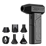
- Powerful Turbo Fan:CAGIWIRU Electric Air Duster have reached speed up to 130000RPM,Can efficient remove dust and debris.With LED light and Three Adjustable Speed to meet with different cleaning tasks.
- With Attached USB C Fasting Charging cable.Charing 3 hours,Enjoy up to 240 minutes of use on the lowest setting, For third speed can use for 25mins.with four charging options to meet with your needs.
- Wide Application: attached 5 different nozzles,making it suitable for a variety of scenes, such as car,pet, pc, keyboards, and other electronic devices. It also serves for office and home clean duster.
- Handy Design & Portable:Cordless air duster electric nozzle is Light weight and small size, design and comfortable to hold and space saving, convenient to carry around. You can put it in your room or inside the car without taking up space.
- Economical &Reusable: Built durable materials with a long-lasting battery,our CAGIWIRU air duster it's a sustainable alternative to disposable air cans.
PSMA PET/CT
In PSMA PET/CT, PET records tracer uptake and CT supplies structural X-ray images. The CT data help localize a focus of uptake, show its relationship to organs and bones, and support PET attenuation correction. A PET/CT examination may use a low-dose CT for attenuation correction and anatomic correlation, or it may include a separately optimized diagnostic CT protocol. A low-dose companion CT should not automatically be described as a replacement for a full diagnostic CT.
The joint EANM/SNMMI procedure guideline 2.0, published in 2023, provides detailed professional guidance for PSMA-ligand PET/CT. Its routine clinical applications include several important prostate-cancer scenarios, described below.
PSMA PET/MRI
PSMA PET/MRI combines the same PET tracer-distribution information with anatomy obtained from magnetic resonance imaging. The guideline defines PET/MRI as an integrated or multimodality system in which PET and MRI can be acquired sequentially or simultaneously while the patient remains in the same position.
Free tools Windows power users keep installed
One-click scans. No signup required.
Published reports describe PET/MRI use for initial staging and biochemical recurrence. However, the joint EANM/SNMMI guideline explicitly excludes PET/MRI protocols from its scope. That means the document cannot be used to claim that PET/MRI is universally more accurate, safer, or preferable to PET/CT. The two combinations answer overlapping but not identical imaging needs, and local protocols differ.
Comparison at a glance
| Term | What provides molecular information? | What provides anatomy? | What the evidence and guidance establish |
|---|---|---|---|
| PSMA PET | PET detection of a radiolabeled PSMA-targeting ligand | Not specified by the term; anatomic imaging is normally paired with PET | Describes the tracer-distribution component, not a complete statement of the accompanying scan |
| PSMA PET/CT | PET tracer uptake | CT, with the protocol ranging from low-dose attenuation-correction CT to a diagnostic CT examination | Joint EANM/SNMMI 2023 guidance addresses professional PET/CT protocols and several established clinical uses |
| PSMA PET/MRI | PET tracer uptake | MRI-derived anatomy, acquired sequentially or simultaneously with PET | Reported in some initial-staging and recurrence settings; PET/MRI protocols are outside the joint guideline’s scope |
How PSMA uptake should be interpreted
PSMA PET shows where the tracer accumulates; it does not directly display a pathology result. Normal organs can have physiologic uptake, and uptake can also occur in some non-malignant conditions or cancers other than prostate cancer. The U.S. Food and Drug Administration (FDA) notes that the Pylarify tracer can bind in other cancers and in certain benign conditions, creating potential interpretation errors.
Rank #2

- Screen Cleaner is Your Screen’s New Bestfriend! - EVEO is proud to present a screen cleaner that’s perfectly suited for your TV. Meet your new screen cleaner, computer screen cleaner, laptop cleaner, iPad cleaner, Macbook,tv cleaner or any other electronic device. Our screen cleaner spray and wipe along with the included plush microfiber cloth, easily removes dust, fingerprints & other smudges on the screen’s surface. Clean effectively for a streak-free screen
- Gentle on your Screens - Our screen cleaner spray and wipes is compatible to be used even for the most sensitive LCD, LED, CRT, and OLED screens in proper use. Be at ease knowing that this product is compatible with all the major brands: LG, SONY, Samsung, Sharp, iPad, iPhone, Android screens, Kindle, PC monitors - we’ve got you covered! Can be used as LCD screen cleaner, tv cleaner, smart tv screen cleaner, and more
- Premium Super Soft Plush Microfiber Cloth - Included in this efficient screen cleaning kit. This screen cleaner spray and wipe has our signature plush microfiber cloth specially designed to comply with screens and monitors and leave them streak-free. Has been shown to effectively absorbs stubborn stains, zero streaks, and no fiber shedding. Always use this together with our screen cleaner for the best results
- Easy-to-Use Screen Cleaner Kit + microfiber cleaning cloth - The EVEO Screen Cleaning Kit will easily be your new favorite tool when it comes to taking care of the things you love most, quick and easy solution for a streak-free screen. Simply screw on the sprayer gun on the screen cleaner bottle. Spray the cleaning liquid onto the microfiber cloth, then wipe the screen until desired cleanliness & shine is achieved.
- Quality Screen Care Kit Worth Purchasing - Enjoy a convenient Screen Cleaner anytime, anywhere you need it. Your satisfaction is our top priority. We stand behind the quality of our products and that’s why, you shouldn’t be worrying at all. We manufacture the Best Screen Cleaner. and can be used for laptop cleaning kit, laptop screen cleaner, tv screen cleaner for smart tv. MacBook screen cleaner, monitor cleaner or screen cleaner spray for electronic devices with microfiber cleaning cloth
Expression is not identical in every tumor. Some prostate-cancer deposits express less PSMA than others, and very small lesions may not accumulate enough tracer to be resolved. SNMMI’s patient guidance gives a practical limitation: tumors smaller than about 0.5 cm may not appear on the scan. Consequently, a bright focus is not automatically cancer, and a negative study does not prove that no disease exists.
“No imaging test is perfect.”
That statement comes from SNMMI’s patient guide, which discusses both false-positive and false-negative findings. The treating team must interpret the images and report together with PSA history, examination, pathology, prior imaging and the reason the scan was ordered.
When PSMA PET/CT is used
The EANM/SNMMI procedure guideline 2.0 supports PSMA-ligand PET/CT for defined clinical questions rather than as a general screening test for people without a specific prostate-cancer indication.
Initial staging
For newly diagnosed disease, PSMA PET/CT can be used when the purpose is to stage the cancer, particularly when the disease is considered high risk and the result could change treatment. In the phase III, multicenter randomized ProPSMA trial, as reported by the 2023 guideline, gallium-68 PSMA-11 PET/CT had 27% greater accuracy than CT plus bone scanning for initial staging of people with high-risk prostate cancer. That percentage applies to the trial population and its specific comparison; it is not a universal estimate for PET/CT versus PET/MRI.
Persistent or recurrent disease
After treatment, a rising or persistently detectable PSA can prompt imaging to locate residual or recurrent disease. The guideline lists localization of biochemical persistence or recurrence among the routine PET/CT indications it addresses. The scan may identify disease in the prostate bed, lymph nodes, bones or other sites, but each finding still requires clinical correlation.
Rank #3

- 【Large size】:Measuring 800 x 300 mm/ 31. 5×12 inches, which is wider and taller than others. Large mouse pad can perfectly fit your keyboard and mouse of any size, and there is also ample space for peripherals such as phones, tablets, etc.
- 【LED backlight mouse pad】:Blade Hawks RGB mouse pad provides 7 static modes and 7 dynamic modes will give you the options you are looking for. Power-off memory function remembers the last lighting mode you selected. The super glow fiber Chroma will give you a colorful gaming setup that you want.
- 【Anti-slip rubber base】:Made from Natural rubber, this gaming mousepad will firmly grip your desktop, allowing you to enjoy the ultimate precision in the games you are most passionate about. It also comes with a texture that enhances this ability even further.
- 【Smooth and waterproof surface】: Micro-textured cloth surface, for extremely precise mouse control and a smooth movement. When liquid splashes on the gaming mouse pad, it forms water droplets and slides down. Will not delay your work or gameplay.
- 【Plug and play】: This mouse and keyboard pad is powered by USB, plug and play, no driver required. One button control light modes. Press one time to change the lighting mode, press twice to change the brightness, press and hold about 3 seconds turn ON/OFF.
Conventionally non-metastatic castration-resistant prostate cancer
The guideline includes localization in selected patients whose disease is considered conventionally non-metastatic castration-resistant prostate cancer. “Selected” matters: the decision depends on the patient’s prior imaging, treatment history and the question the clinician needs answered.
Planning PSMA-directed radioligand therapy
PSMA PET/CT can stage disease before PSMA-directed radioligand therapy. In that setting, the scan helps determine whether lesions show sufficient target expression under the treatment program’s criteria. It is a treatment-selection and staging examination, not a guarantee that every deposit will respond.
Other and emerging applications
The guideline also discusses possible roles in biopsy guidance and imaging of metastatic disease, while emphasizing uncertainty for some emerging uses. A use being discussed in a guideline is not the same as a universal recommendation for every patient or health system.
What the reported studies show—and what they do not show
ProPSMA: 27% greater accuracy in a defined staging comparison
The ProPSMA result compares gallium-68 PSMA-11 PET/CT with conventional CT and bone scanning in people with high-risk prostate cancer undergoing initial staging. It demonstrates the value of PSMA PET/CT for that population and decision. It does not compare PET/CT with PET/MRI, does not apply to every risk group, and should not be presented as a general superiority percentage.
Pelvic lymph-node micrometastases can be missed
Across two phase II/III studies summarized by the guideline, pelvic-node specificity was at least 95%, while sensitivity was in the 40% range. High specificity means a positive result can be useful when the finding is correctly characterized; sensitivity in that range means a negative scan cannot exclude microscopic cancer in pelvic lymph nodes. Surgical, pathologic or other clinical information may still be needed.
Quick wins for a faster PC:
Fix the driver behind crashes, sound loss and screen glitchesFind Drivers →Repair Windows errors before they cause bigger problemsFix Now →Rank #4

- 【Softer and More Comfortable】Vaydeer wrist rest has unique diamond pattern, which is the combination of softness and aesthetics. The materials of wrist rest are improved into higher quality memory foam and covered with silky smooth lycra. The computer wrist rest makes you as comfortable and cushiony as like rest your wrists on clouds.
- 【Ergonomic Wrist Saver】The wrist rests for keyboard and mouse comes with a 17.32×3.15×0.83 inch keyboard wrist pad and a 5.94×3.15×0.83 inch mouse wrist support. Based on ergonomic design, the unique concave shape is the perfect fit for your wrist joints. The wrist rest pad fits most computer keyboards and laptops, improve hand and wrist posture, release your wrist and arm stress.
- 【Non-Slip Rubber Bottom】Featuring an anti-skid silicone base on the bottom, this wrist keyboard support stays firmly in place on your desk, preventing the padding from sliding around, ensuring stable and consistent wrist support during extended computer sessions.
- 【Better Experience & Pain Relief】Our keyboard arm rest is beneficial to alleviate the soreness caused by direct contact and friction between your arm and a hard desk surface, reducing the risk of wrist fatigue or carpal tunnel. The soft texture of memory foam can evenly distribute the pressure around your wrists and provide good support with just enough give.
- 【Helpful in Multiple Scenarios】Whether you're working, studying, writing, typing, gaming, this keyboard and mouse rest combo is an essential accessory to add comfort and support to your hands and wrists. It’s also a great gift for men, women, family, friend, coworker, gamer, teacher, etc.
PRIMARY: adding PSMA PET to multiparametric MRI for biopsy guidance
In the prospective PRIMARY study, adding gallium-68 PSMA-11 PET to multiparametric MRI improved the negative predictive value from 72% to 91% and sensitivity from 83% to 97% for clinically significant prostate cancer. The guideline uses this evidence to support combining PSMA PET with multiparametric MRI for biopsy guidance. These results concern an added PET-plus-multiparametric-MRI approach in that setting; they do not prove that a whole-body PSMA PET/MRI examination is always superior to PSMA PET/CT.
FDA Pylarify study after definitive treatment
In an FDA-reviewed study of 208 people with rising PSA after definitive treatment, whose baseline conventional imaging did not show definite spread, Pylarify PET/CT detected at least one positive lesion in 60% of participants. Among patients with correlative pathology, imaging or serial PSA data, the FDA reported confirmation of positive readings in an estimated 85% to 87%, depending on the reader. Those percentages belong to that cohort and verification process; they are not a universal detection rate for all PSMA PET examinations.
How a typical examination works
- Clinical order and protocol selection: The treating clinician states the question—such as initial staging, suspected recurrence or therapy eligibility—and the imaging service selects an appropriate tracer and PET/CT or PET/MRI protocol.
- Tracer injection: A radiotracer is administered through an intravenous line. The exact agent, activity and approval status vary by jurisdiction and facility. SNMMI notes that several FDA-approved PSMA radiotracers perform similarly, but availability and regulatory approvals are geographic and time-sensitive.
- Uptake period: The patient generally waits about an hour after injection so the tracer can distribute. This is an estimate from SNMMI’s patient guide; local timing can differ.
- Image acquisition: A typical scan takes about 30 minutes, according to the same guide. PET/CT adds CT acquisition; PET/MRI adds MRI sequences. The patient may need to remain still, and the exact sequence length depends on the protocol.
- Specialist interpretation: An imaging specialist reviews tracer distribution, the accompanying anatomy and relevant prior studies, then issues a report. The care team combines that report with PSA values, pathology, treatment history and examination findings.
Limitations, false positives and false negatives
Small or low-expression tumors
Lesions below roughly 0.5 cm may be below the scan’s ability to resolve, and tumors with limited PSMA expression may be less conspicuous. A negative scan therefore cannot exclude every focus of disease.
Pelvic-node micrometastases
The phase II/III evidence summarized above is especially important for surgical planning: sensitivity in the 40% range means microscopic nodal disease can remain after a negative PET/CT result.
Benign and non-prostate uptake
Normal physiologic uptake and uptake associated with benign conditions or other cancers can mimic prostate-cancer deposits. Radiologists and nuclear-medicine physicians assess the pattern, location and CT or MRI appearance rather than treating every focus as malignant.
Best Value

- 【No Drill Mounted】Cable management tray can be assembled in as little as 3 minutes or less-simply. Side clips easily clampe the edge of desk, pass through the holes on the sides, and secure excess cables with baskets. Desk cable management supports inward or outward installation to meet your needs in different scenarios, and provides you with great convenience when collecting and organizing wires.
- 【Rubber Pad Protects Desk】Wire organizer under desk built-in soft rubber pads on the clamp protect your desk from scratches, so you will not damage your furniture or office.
- 【Sturdy and Beautiful】Cord management under desk is made of high-quality carbon steel and holds about 15 lbs. The wire basket has 2 holes on each side of the tray to enable your cables to pass freely. Cable baskets are suitable for desk thickness use from 0.4" to 2.4".
- 【Space Saving & Keep Organized】Desk cable management tray holds all your power cords, outlet strips, USB hubs and computer transformers hidden from your feet, and the data cables will not enter the vacuum cleaner to keep your desk clean and tidy. Desk wire organizer is also suitable for use in the kitchen and outdoors.
- 【Reduce Safety Risks】Cable tray under desk keeps all plugs away from your kids, no more worry about tripping. Using 2 holes wire tray saves extra space, helps you easily cabling and wire protection. The mesh design is not easy to accumulate dust, but avoids safety accidents caused by messy wires.
Technical and protocol variation
A low-dose CT acquired for attenuation correction is not equivalent to a dedicated diagnostic CT. PET/MRI examinations can also differ in whether PET and MRI are simultaneous or sequential and which MRI sequences are included. Comparing reports without checking the protocol can create a misleading impression of test performance.
Choosing between PET/CT and PET/MRI
There is no evidence-supported winner for every clinical question. A practical decision uses the following sequence.
- Define the question. Ask whether the goal is initial staging, locating recurrence, assessing a specific lesion, guiding a biopsy or evaluating eligibility for PSMA-directed radioligand therapy.
- Identify the anatomic information needed. CT may be adequate for structural localization and attenuation correction in many PET/CT protocols. MRI may be selected when the clinical team needs MRI-based soft-tissue or pelvic anatomy under a local protocol.
- Check the exact protocol. Confirm whether CT is low-dose or diagnostic, which MRI sequences are planned, whether contrast is used, and whether PET and MRI are acquired sequentially or simultaneously.
- Account for patient suitability. The service must consider the patient’s ability to remain still, prior imaging, devices or other factors that affect MRI or CT, and the availability of an experienced interpreting team.
- Use local expertise and access. A technically suitable examination performed and interpreted by a team familiar with PSMA imaging may be more useful than an unavailable or unfamiliar alternative. The joint guideline’s PET/CT recommendations should not be silently transferred to PET/MRI.
- Discuss how the result will change management. If a positive or negative scan would not alter treatment, the clinician may choose a different test or defer imaging. Decisions belong with the treating team, not with a generic modality ranking.
Questions to ask before scheduling
- What specific clinical question is the scan intended to answer?
- Which PSMA tracer will be used, and is it approved or available in this jurisdiction?
- Is the CT component low-dose for attenuation correction, diagnostic CT, or both?
- If PET/MRI is proposed, which MRI sequences and contrast protocol are included?
- How will the result be reconciled with PSA, pathology and previous scans?
- Could a negative result still miss microscopic nodal or very small disease?
- Who will interpret the study, and will the prior images and reports be available?
How to read a PSMA PET report without overreading it
Start with the indication and tracer named in the report. Then identify the locations of uptake and whether the accompanying CT or MRI shows a corresponding structural abnormality. A report may describe physiologic uptake, indeterminate findings, probable disease or findings that require correlation; those categories are not interchangeable.
Pay attention to the impression and to recommendations for comparison, MRI, biopsy, follow-up or other testing. A statement that no definite PSMA-avid disease was identified means no convincing abnormal uptake was seen under that protocol; it does not eliminate microscopic nodal disease or every lesion below the technique’s resolution.
Likewise, a positive focus is an imaging finding, not a pathology result. The treating clinician may use PSA kinetics, prior treatment, biopsy results, anatomic appearance and changes over time to decide whether additional confirmation or treatment is appropriate.
Bottom line
PSMA PET is the molecular map of radiotracer uptake. PET/CT adds CT anatomy and is the combination covered by established joint EANM/SNMMI guidance for several prostate-cancer decisions. PET/MRI adds MRI anatomy and has reported applications, but its protocols were outside that guideline’s scope. The clinically correct choice is the one that answers the patient’s specific question with an appropriate protocol and qualified interpretation—not a blanket claim that one combination is always best.
Quick Recap
Product prices and availability are accurate as of the date/time indicated and are subject to change. Any price and availability information displayed on Amazon at the time of purchase will apply.
Recommended Free Tools

