
iTechGuides is reader-supported. When you buy through links on our site, we may earn an affiliate commission. As an Amazon Associate I earn from qualifying purchases. Learn more
There is no universal winner. For rapid, long-duration live imaging of developing mouse embryos, light-sheet microscopy is often the best fit. Confocal microscopy remains valuable when high-resolution optical sectioning and fluorescence detail matter. MRI is the practical alternative when non-destructive, whole-volume or in-utero imaging is more important than cellular-scale detail. Embryo stage, live versus fixed state, labels, optical access, depth, field of view and throughput should determine the choice.
What each method actually does
The three techniques form different compromises between illumination, access, resolution, speed and specimen handling. Treating them as interchangeable can produce an experiment that is technically impressive but unable to answer the biological question.
| Method | Signal and acquisition | Best-supported strengths | Main constraints |
|---|---|---|---|
| Light-sheet | A thin plane is illuminated from a direction separate from the detection objective. | Fast volumetric acquisition, lower unnecessary illumination, and extended live imaging in suitable preparations. | Requires specialized mounting, optical access and environmental control; performance varies by implementation. |
| Confocal | A focused laser spot is scanned through the sample to build an image by optical section. | Detailed fluorescence imaging and optical sectioning with widely established workflows. | Scanning exposes the sample during acquisition; speed, bleaching and depth depend on scan strategy and specimen properties. |
| MRI | Magnetic-resonance signals are reconstructed into a three-dimensional volume. | Non-destructive coverage of intact specimens and access to settings where optical access is difficult, including in-utero work described in mouse phenotyping literature. | Micro-MRI generally has less spatial resolution than optical methods for detailed embryo phenotyping; signal-to-noise and coil design are critical. |
These are modality-level descriptions, not guarantees for every instrument. Objective numerical comparisons belong to the particular system, sample and acquisition settings that produced them.
How light-sheet microscopy works for embryos
In light-sheet microscopy, illumination and detection are arranged on separate optical paths. A sheet of light intersects one plane of the embryo while the detection objective records fluorescence from that plane. The system then moves through the specimen or changes the illuminated plane to build a volume.
#1 Best Overall
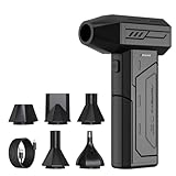
- Powerful Turbo Fan:CAGIWIRU Electric Air Duster have reached speed up to 130000RPM,Can efficient remove dust and debris.With LED light and Three Adjustable Speed to meet with different cleaning tasks.
- With Attached USB C Fasting Charging cable.Charing 3 hours,Enjoy up to 240 minutes of use on the lowest setting, For third speed can use for 25mins.with four charging options to meet with your needs.
- Wide Application: attached 5 different nozzles,making it suitable for a variety of scenes, such as car,pet, pc, keyboards, and other electronic devices. It also serves for office and home clean duster.
- Handy Design & Portable:Cordless air duster electric nozzle is Light weight and small size, design and comfortable to hold and space saving, convenient to carry around. You can put it in your room or inside the car without taking up space.
- Economical &Reusable: Built durable materials with a long-lasting battery,our CAGIWIRU air duster it's a sustainable alternative to disposable air cans.
Because regions outside the selected plane are not continuously illuminated, the method can reduce bleaching and photodamage compared with scanning approaches. That geometry is especially useful for time-lapse experiments in which a developing embryo must be imaged repeatedly for hours. The 2009 selective-plane-illumination literature identifies rapid acquisition and reduced exposure as central advantages, while later developmental reviews emphasize the value for extended live imaging.
What must be solved before imaging
- Mounting: The embryo must be held in a position compatible with both the illumination sheet and detection objective. Mounting geometry can limit the angles available for multiview imaging.
- Physiology: Ex-utero imaging requires control of temperature, gas exchange, medium and mechanical stability. A fast camera cannot compensate for developmental arrest caused by poor environmental control.
- Optical access: Scattering, pigmentation, tissue thickness and the selected label determine how far the sheet and detection path can reach.
- Data handling: Multiview acquisition and computational fusion can produce very large three-dimensional datasets. Storage, registration and segmentation should be planned before the first time point.
Protocols published in 2022 cover living, fixed and cleared specimens, so light-sheet is not limited to one preparation type. Clearing can improve access to fixed embryos, but it changes tissue properties and is not a substitute for a viable live preparation.
Rank #2

- Screen Cleaner is Your Screen’s New Bestfriend! - EVEO is proud to present a screen cleaner that’s perfectly suited for your TV. Meet your new screen cleaner, computer screen cleaner, laptop cleaner, iPad cleaner, Macbook,tv cleaner or any other electronic device. Our screen cleaner spray and wipe along with the included plush microfiber cloth, easily removes dust, fingerprints & other smudges on the screen’s surface. Clean effectively for a streak-free screen
- Gentle on your Screens - Our screen cleaner spray and wipes is compatible to be used even for the most sensitive LCD, LED, CRT, and OLED screens in proper use. Be at ease knowing that this product is compatible with all the major brands: LG, SONY, Samsung, Sharp, iPad, iPhone, Android screens, Kindle, PC monitors - we’ve got you covered! Can be used as LCD screen cleaner, tv cleaner, smart tv screen cleaner, and more
- Premium Super Soft Plush Microfiber Cloth - Included in this efficient screen cleaning kit. This screen cleaner spray and wipe has our signature plush microfiber cloth specially designed to comply with screens and monitors and leave them streak-free. Has been shown to effectively absorbs stubborn stains, zero streaks, and no fiber shedding. Always use this together with our screen cleaner for the best results
- Easy-to-Use Screen Cleaner Kit + microfiber cleaning cloth - The EVEO Screen Cleaning Kit will easily be your new favorite tool when it comes to taking care of the things you love most, quick and easy solution for a streak-free screen. Simply screw on the sprayer gun on the screen cleaner bottle. Spray the cleaning liquid onto the microfiber cloth, then wipe the screen until desired cleanliness & shine is achieved.
- Quality Screen Care Kit Worth Purchasing - Enjoy a convenient Screen Cleaner anytime, anywhere you need it. Your satisfaction is our top priority. We stand behind the quality of our products and that’s why, you shouldn’t be worrying at all. We manufacture the Best Screen Cleaner. and can be used for laptop cleaning kit, laptop screen cleaner, tv screen cleaner for smart tv. MacBook screen cleaner, monitor cleaner or screen cleaner spray for electronic devices with microfiber cleaning cloth
What confocal microscopy contributes
Confocal microscopy forms an image by scanning a focused laser spot across the sample and rejecting out-of-focus light during detection. Stacking optical sections gives a three-dimensional reconstruction with strong fluorescence contrast and familiar quantitative workflows.
Recommended Free Tools
When confocal is the better fit
- You need established optical-sectioning methods for a fluorescent reporter, immunolabel or stained fixed embryo.
- The region of interest is relatively small, so a full-embryo rapid volume is unnecessary.
- You need the flexibility of conventional objectives, sample holders and analysis pipelines already available in the laboratory.
- You are comparing a new preparation with a large body of existing confocal data.
Confocal acquisition is inherently a scanning process. In the comparison summarized by Keller and Stelzer, the scanned sample receives illumination during volume acquisition, whereas the light-sheet implementation restricted illumination more closely to the observed plane. That difference can matter for repeated live imaging, but it does not make every confocal experiment unsuitable: scan speed, laser power, pixel dwell time, z-step, objective numerical aperture and specimen sensitivity all affect the outcome.
Rank #3

- 【Large size】:Measuring 800 x 300 mm/ 31. 5×12 inches, which is wider and taller than others. Large mouse pad can perfectly fit your keyboard and mouse of any size, and there is also ample space for peripherals such as phones, tablets, etc.
- 【LED backlight mouse pad】:Blade Hawks RGB mouse pad provides 7 static modes and 7 dynamic modes will give you the options you are looking for. Power-off memory function remembers the last lighting mode you selected. The super glow fiber Chroma will give you a colorful gaming setup that you want.
- 【Anti-slip rubber base】:Made from Natural rubber, this gaming mousepad will firmly grip your desktop, allowing you to enjoy the ultimate precision in the games you are most passionate about. It also comes with a texture that enhances this ability even further.
- 【Smooth and waterproof surface】: Micro-textured cloth surface, for extremely precise mouse control and a smooth movement. When liquid splashes on the gaming mouse pad, it forms water droplets and slides down. Will not delay your work or gameplay.
- 【Plug and play】: This mouse and keyboard pad is powered by USB, plug and play, no driver required. One button control light modes. Press one time to change the lighting mode, press twice to change the brightness, press and hold about 3 seconds turn ON/OFF.
Confocal limitations in whole embryos
Optical scattering and absorption limit depth. A large embryo may require compromises in z-step, field of view, objective working distance or detector settings. Increasing laser power can recover signal while accelerating bleaching or phototoxicity. If the biological question requires a complete embryo volume at many time points, acquisition time and light dose should be estimated from a pilot stack rather than assumed from a single-frame specification.
What MRI can and cannot answer
MRI detects magnetic-resonance signals and reconstructs them into a three-dimensional volume without optical illumination. That makes it non-destructive and attractive when the specimen must remain intact or when the uterus prevents direct optical access. The mouse embryo phenotyping review describes in-utero use cases in which avoiding optical exposure and surgical access is a decisive advantage.
Rank #4

- 【Softer and More Comfortable】Vaydeer wrist rest has unique diamond pattern, which is the combination of softness and aesthetics. The materials of wrist rest are improved into higher quality memory foam and covered with silky smooth lycra. The computer wrist rest makes you as comfortable and cushiony as like rest your wrists on clouds.
- 【Ergonomic Wrist Saver】The wrist rests for keyboard and mouse comes with a 17.32×3.15×0.83 inch keyboard wrist pad and a 5.94×3.15×0.83 inch mouse wrist support. Based on ergonomic design, the unique concave shape is the perfect fit for your wrist joints. The wrist rest pad fits most computer keyboards and laptops, improve hand and wrist posture, release your wrist and arm stress.
- 【Non-Slip Rubber Bottom】Featuring an anti-skid silicone base on the bottom, this wrist keyboard support stays firmly in place on your desk, preventing the padding from sliding around, ensuring stable and consistent wrist support during extended computer sessions.
- 【Better Experience & Pain Relief】Our keyboard arm rest is beneficial to alleviate the soreness caused by direct contact and friction between your arm and a hard desk surface, reducing the risk of wrist fatigue or carpal tunnel. The soft texture of memory foam can evenly distribute the pressure around your wrists and provide good support with just enough give.
- 【Helpful in Multiple Scenarios】Whether you're working, studying, writing, typing, gaming, this keyboard and mouse rest combo is an essential accessory to add comfort and support to your hands and wrists. It’s also a great gift for men, women, family, friend, coworker, gamer, teacher, etc.
Strengths of micro-MRI
- Whole-volume coverage without physically sectioning the embryo.
- Compatibility with intact-animal or in-utero questions when the protocol and hardware support them.
- No fluorophore bleaching or optical scattering in the conventional sense.
- Ability to repeat non-destructive measurements when temporal and physiological constraints permit.
Resolution and hardware trade-offs
The same phenotyping review cautions that micro-MRI does not reach the spatial resolution needed for detailed mouse-embryo phenotyping that optical methods can provide. MRI can show overall anatomy and volume relationships, but it should not be selected on the assumption that it will resolve every cellular or fine fluorescent feature visible in a microscope.
Signal-to-noise ratio (SNR) is a practical design variable. A 2010 comparative mouse-embryo MR-microscopy study reported 3.3 times as much SNR for an individual-coil acquisition as for a shared-coil acquisition under equivalent conditions. The individual-coil arrangement was harder and more expensive to implement, constrained receiver channels and reduced the convenience of handling multiple specimens. That figure is evidence for a specific coil comparison, not a general MRI performance guarantee.
Best Value

- 【No Drill Mounted】Cable management tray can be assembled in as little as 3 minutes or less-simply. Side clips easily clampe the edge of desk, pass through the holes on the sides, and secure excess cables with baskets. Desk cable management supports inward or outward installation to meet your needs in different scenarios, and provides you with great convenience when collecting and organizing wires.
- 【Rubber Pad Protects Desk】Wire organizer under desk built-in soft rubber pads on the clamp protect your desk from scratches, so you will not damage your furniture or office.
- 【Sturdy and Beautiful】Cord management under desk is made of high-quality carbon steel and holds about 15 lbs. The wire basket has 2 holes on each side of the tray to enable your cables to pass freely. Cable baskets are suitable for desk thickness use from 0.4" to 2.4".
- 【Space Saving & Keep Organized】Desk cable management tray holds all your power cords, outlet strips, USB hubs and computer transformers hidden from your feet, and the data cables will not enter the vacuum cleaner to keep your desk clean and tidy. Desk wire organizer is also suitable for use in the kitchen and outdoors.
- 【Reduce Safety Risks】Cable tray under desk keeps all plugs away from your kids, no more worry about tripping. Using 2 holes wire tray saves extra space, helps you easily cabling and wire protection. The mesh design is not easy to accumulate dust, but avoids safety accidents caused by messy wires.
Which method fits your experiment?
| Experimental priority | First method to evaluate | Reasoning |
|---|---|---|
| Long live time-lapse with many three-dimensional time points | Light-sheet | Plane illumination can reduce exposure and support rapid volumes, provided mounting and physiology are controlled. |
| High-detail fluorescence in a limited region or fixed specimen | Confocal | Optical sectioning and established fluorescence workflows are often sufficient without whole-embryo throughput. |
| Intact or in-utero three-dimensional coverage | MRI | Non-optical access avoids the need to expose the embryo directly, with a resolution trade-off. |
| Cleared fixed embryo requiring large-volume optical imaging | Light-sheet | Clearing and multiview acquisition can cover large specimens, subject to protocol and instrument compatibility. |
| Direct comparison with an existing confocal dataset | Confocal or a deliberately cross-calibrated light-sheet workflow | Matching labels, sampling and analysis may matter more than nominal acquisition speed. |
For a choice between light-sheet and confocal, ask first whether the experiment is limited by light dose and time or by optical detail in a smaller region. For a choice between optical imaging and MRI, ask whether direct access to the embryo is possible and whether the required resolution is anatomical or cellular.
Independent reader supportYour contribution helps us test, update, and keep practical guides available for everyone.Practical workflow: planning a light-sheet experiment
- Define the biological endpoint. Specify the developmental window, number of time points, required field of view, z-resolution and fluorescent channels.
- Choose live, fixed or cleared preparation. Live work demands environmental control; fixed or cleared work allows different mounting and labeling choices.
- Test the mount with an unlabeled or low-value specimen. Confirm that the embryo remains centered as the stage moves and that the detection objective can reach the intended planes.
- Run a low-dose pilot. Measure signal, bleaching, drift and developmental health before committing to a long time-lapse.
- Plan multiview acquisition and registration. Decide whether multiple angles are needed and allocate time for fusion, deconvolution or segmentation.
- Record acquisition metadata. Keep objective, sheet thickness or illumination settings, exposure, z-step, time interval, temperature and medium with every dataset.
Practical workflow: planning a confocal experiment
- Set the optical sampling first. Select objective, pixel size and z-step according to the smallest feature that must be resolved, not simply the camera’s maximum resolution.
- Measure the usable depth. Acquire a test stack through the embryo and note where scattering or signal loss makes quantitative interpretation unreliable.
- Minimize dose during the pilot. Start with the lowest laser power and shortest dwell time that produce an analyzable signal; increase only when the measurement requires it.
- Choose scan mode deliberately. A smaller region, fewer channels or a coarser preview stack can reduce exposure before the final high-resolution acquisition.
- Validate live compatibility. For time-lapse work, compare developmental progression in imaged and control embryos under the same environmental conditions.
Practical workflow: planning an MRI experiment
- State the access requirement. Decide whether the study needs an intact, in-utero or ex-utero volume and whether repeated non-destructive measurements are necessary.
- Define the minimum anatomical resolution. If the endpoint is fine phenotyping, verify that the proposed micro-MRI protocol can resolve it before investing in a large cohort.
- Choose coil architecture. Compare individual- and shared-coil options for SNR, receiver-channel availability, specimen throughput, implementation difficulty and cost.
- Benchmark SNR on representative embryos. Use the same geometry and acquisition conditions that will be used for the study; published SNR ratios cannot be transferred automatically between scanners.
- Plan motion and physiology controls. In-utero and live-animal measurements require an appropriate preparation, monitoring and timing plan so motion does not erase the advantage of whole-volume coverage.
Performance figures: useful context, not universal specifications
Historical light-sheet literature provides striking system-specific measurements. Keller and Stelzer’s 2009 review reported up to 50 times higher imaging speeds, 10–100 times higher signal-to-noise ratios and at least three orders of magnitude less light energy for its digital scanned laser light-sheet implementation than the compared confocal and two-photon fluorescence systems. Those values describe the systems and conditions discussed in that review; they should not be quoted as guaranteed advantages for every modern instrument or embryo.
Tomer and colleagues reported an acquisition rate of 175 million voxels per second for the SiMView simultaneous multiview system in 2012. That number demonstrates what a particular architecture achieved, not a standard rate for all light-sheet microscopes. Throughput still depends on volume size, number of views, channels, camera mode, z-step and processing time.
Quick wins for a faster PC:
Scan for outdated or missing drivers - takes under a minuteDriver Scan →Clear out junk files and repair common Windows errorsFree Scan →Fix the driver behind crashes, sound loss and screen glitchesFind Drivers →The 3.3-fold MRI SNR result likewise belongs to the individual-versus-shared-coil comparison described above. It is most useful as a reminder that hardware configuration can change data quality as much as the nominal modality.
Common failure modes and fixes
- Embryo drifts during a light-sheet time-lapse: Improve mechanical support and verify that the mount remains compatible with medium exchange and developmental movement.
- Rapid bleaching or developmental damage: Reduce exposure, laser power and unnecessary channels; shorten the illuminated volume or time interval where the endpoint allows.
- Uneven brightness between light-sheet views: Check alignment, sheet uniformity and view registration before interpreting intensity differences biologically.
- Confocal stack loses signal with depth: Test a lower-scattering preparation, adjust labeling and objective choice, or restrict the claim to the reliably sampled depth.
- Confocal time-lapse changes embryo development: Compare imaged and un imaged controls and reduce scan burden before increasing laser power.
- MRI volume looks anatomically complete but lacks phenotyping detail: Revisit the minimum feature size; whole-volume coverage does not imply cellular-scale resolution.
- MRI SNR is inadequate: Evaluate coil geometry, receiver channels, averaging and specimen positioning rather than assuming that longer scans alone will solve the problem.
- Large datasets cannot be analyzed in time: Define storage, backup, registration and segmentation procedures before acquisition and retain raw data with metadata.
A decision checklist
- Is the embryo live for an extended period, or can it be fixed?
- Must the experiment reach the embryo directly, or is intact/in-utero access essential?
- Is the endpoint cellular fluorescence, tissue architecture or gross anatomy?
- How many three-dimensional volumes and time points are required?
- What bleaching, phototoxicity and motion can the specimen tolerate?
- Can the available mounting, environmental-control, coil and receiver hardware support the intended protocol?
- Will the resulting volume be large enough to require multiview registration, segmentation or specialized storage?
Bottom line
Choose light-sheet when fast, gentle, repeated volumetric imaging is the central requirement and you can engineer suitable mounting and environmental control. Choose confocal when established optical sectioning and detailed fluorescence in a manageable volume outweigh whole-embryo speed. Choose MRI when non-destructive three-dimensional access, especially in-utero or intact-specimen coverage, is more important than the finest optical detail. Validate the decision with a pilot on the actual embryo stage, label, geometry and hardware: the published speed, SNR and voxel-rate figures are benchmarks for named systems, not universal promises.
Quick Recap
Product prices and availability are accurate as of the date/time indicated and are subject to change. Any price and availability information displayed on Amazon at the time of purchase will apply.

