
Quick wins for a faster PC:
Fix the driver behind crashes, sound loss and screen glitchesFind Drivers →Repair Windows errors before they cause bigger problemsFix Now →iTechGuides is reader-supported. When you buy through links on our site, we may earn an affiliate commission. As an Amazon Associate I earn from qualifying purchases. Learn more
A defensible microscopy submission lets a reader trace every published or measured image back to the microscope’s unaltered output. Keep the original files, work from copies, record the acquisition context, log each consequential operation in order, and disclose any change that could affect interpretation. Put workflow-wide information in Methods and figure-specific details in legends, then apply the destination journal’s current file, source-data, and retention rules.
The goal is not to ban image processing. It is to make processing reproducible and prevent a display choice from being mistaken for a biological finding. The same record should support your own reanalysis months later, a reviewer’s questions, and an editor’s request for source data.
What a complete record must establish
Your documentation should answer five questions without requiring a reader to guess:
The Tool Desk
Outbyte PC Repair FREERepair Windows errors before they cause bigger problemsFix Now →Outbyte Driver Updater FREEScan for outdated or missing drivers - takes under a minuteDriver Scan →- What was acquired? Identify the specimen preparation, stains or fluorochromes, microscope and objective, camera, imaging medium, environmental conditions, acquisition software, file format and bit depth.
- Which original file produced this figure or measurement? Preserve an unaltered source and maintain a traceable relationship between that source, working files and final outputs.
- What software did you use? Name the acquisition package and every post-acquisition program or plugin that materially changed, measured or rendered the data.
- What operations occurred, in what order and for what reason? Describe steps such as deconvolution, registration, 3D reconstruction, rendering, gamma adjustment, thresholding, segmentation and quantitative measurement rather than saying only that images were “processed.”
- Could the operation change what a reader sees or what a number means? State the adjustment, its scope, whether it was applied consistently, and how the result was calibrated or checked.
The community checklist for quantitative light-microscopy publication treats processing and analysis reporting as part of publishing the data, not as an optional software footnote. Institutional guidance from KU Leuven and the Office of Research Integrity (ORI), along with recommendations from the American Society for Biochemistry and Molecular Biology (ASBMB) and Wiley, converges on this traceability approach. Their scopes differ, so the target journal’s instructions remain controlling.
#1 Best Overall
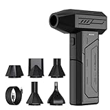
- Powerful Turbo Fan:CAGIWIRU Electric Air Duster have reached speed up to 130000RPM,Can efficient remove dust and debris.With LED light and Three Adjustable Speed to meet with different cleaning tasks.
- With Attached USB C Fasting Charging cable.Charing 3 hours,Enjoy up to 240 minutes of use on the lowest setting, For third speed can use for 25mins.with four charging options to meet with your needs.
- Wide Application: attached 5 different nozzles,making it suitable for a variety of scenes, such as car,pet, pc, keyboards, and other electronic devices. It also serves for office and home clean duster.
- Handy Design & Portable:Cordless air duster electric nozzle is Light weight and small size, design and comfortable to hold and space saving, convenient to carry around. You can put it in your room or inside the car without taking up space.
- Economical &Reusable: Built durable materials with a long-lasting battery,our CAGIWIRU air duster it's a sustainable alternative to disposable air cans.
Preserve originals and build an image lineage
Keep an immutable source set
Copy the microscope’s original files to controlled storage as soon as practical. Do not overwrite them while adjusting brightness, exporting a new format or testing a segmentation method. KU Leuven and ORI both advise retaining the original and manipulating a copy. If the instrument writes metadata, retain that metadata with the file; exporting to a presentation format can discard acquisition information.
Use a read-only location, access-controlled project folder or other mechanism that makes accidental edits difficult. A backup in a separate storage location protects against disk failure, but a backup is not a substitute for an unmodified source. Keep the original bit depth and file format whenever the instrument or journal permits.
Assign a traceable identifier
Give every acquisition a stable identifier that appears in your internal record and maps to the final figure panel or measurement table. A useful record contains:
Free tools Windows power users keep installed
One-click scans. No signup required.
- sample, experiment and acquisition date;
- microscope session or instrument identifier;
- original filename and storage location;
- channel, z-plane or time point where relevant;
- working-copy filename and processing version;
- figure, panel or data-table destination.
Do not rely on a filename such as “final2.tif.” A lineage entry should let another person start with the source and follow each derived file to the submitted output. If one source feeds several panels, record each branch. If a panel combines sources, record every contributing identifier.
Separate analysis data from presentation exports
Keep the data used for measurements distinct from a colorized, downsampled or annotated presentation export. A JPEG or screenshot may be suitable for a submission portal but is generally a poor analysis source because compression and bit-depth conversion can alter intensities. Store the raw or minimally transformed data used for quantification, the parameter file or macro, and the rendered figure as separate artifacts.
Record acquisition context before describing processing
Processing cannot be interpreted without knowing how the signal was acquired. ASBMB’s reporting list includes the following items; Wiley likewise calls for sample, staining, microscope, camera and capture/editing details in Methods or the figure legend.
Rank #2

- Screen Cleaner is Your Screen’s New Bestfriend! - EVEO is proud to present a screen cleaner that’s perfectly suited for your TV. Meet your new screen cleaner, computer screen cleaner, laptop cleaner, iPad cleaner, Macbook,tv cleaner or any other electronic device. Our screen cleaner spray and wipe along with the included plush microfiber cloth, easily removes dust, fingerprints & other smudges on the screen’s surface. Clean effectively for a streak-free screen
- Gentle on your Screens - Our screen cleaner spray and wipes is compatible to be used even for the most sensitive LCD, LED, CRT, and OLED screens in proper use. Be at ease knowing that this product is compatible with all the major brands: LG, SONY, Samsung, Sharp, iPad, iPhone, Android screens, Kindle, PC monitors - we’ve got you covered! Can be used as LCD screen cleaner, tv cleaner, smart tv screen cleaner, and more
- Premium Super Soft Plush Microfiber Cloth - Included in this efficient screen cleaning kit. This screen cleaner spray and wipe has our signature plush microfiber cloth specially designed to comply with screens and monitors and leave them streak-free. Has been shown to effectively absorbs stubborn stains, zero streaks, and no fiber shedding. Always use this together with our screen cleaner for the best results
- Easy-to-Use Screen Cleaner Kit + microfiber cleaning cloth - The EVEO Screen Cleaning Kit will easily be your new favorite tool when it comes to taking care of the things you love most, quick and easy solution for a streak-free screen. Simply screw on the sprayer gun on the screen cleaner bottle. Spray the cleaning liquid onto the microfiber cloth, then wipe the screen until desired cleanliness & shine is achieved.
- Quality Screen Care Kit Worth Purchasing - Enjoy a convenient Screen Cleaner anytime, anywhere you need it. Your satisfaction is our top priority. We stand behind the quality of our products and that’s why, you shouldn’t be worrying at all. We manufacture the Best Screen Cleaner. and can be used for laptop cleaning kit, laptop screen cleaner, tv screen cleaner for smart tv. MacBook screen cleaner, monitor cleaner or screen cleaner spray for electronic devices with microfiber cleaning cloth
| Category | Record | Why it matters |
|---|---|---|
| Sample and preparation | Specimen preparation, fixation or live-imaging conditions, stains and fluorochromes, mounting or imaging medium, temperature and other relevant environmental settings | Signal intensity, background and morphology depend on preparation and environment. |
| Microscope | Make and model; modality where relevant; objective type, magnification and numerical aperture | Optical performance and resolution cannot be inferred from a figure alone. |
| Detector | Camera make and model, detector settings and channel configuration when they affect measurement | Detector response and channel settings influence quantitative values. |
| Acquisition software | Program name and version when available, plus file format and bit depth | The software and encoding determine what metadata and intensity range are retained. |
| Processing software | Each program, plugin or script used after acquisition, with version or release identifier when available | Different implementations can produce different results from the same source. |
| Scale and consistency | Pixel or voxel dimensions, scale bars, and whether compared images were acquired under identical conditions | Readers need a physical reference and a fair basis for comparison. |
Include acquisition settings that materially affect the result, such as exposure, illumination, gain, z-step, time interval or averaging, when your journal or analysis requires them. The cited guidance specifically emphasizes the items in the table; add any instrument parameter needed to reproduce your measurement rather than listing every irrelevant control-panel setting.
Create a processing log that another person can follow
Describe operations in sequence
Write the workflow as an ordered chain from source to output. For each step, state the operation, the input, the important parameters, the reason for using it and the output. For example, “deconvolved” is incomplete; identify the algorithm or software, the measured or theoretical point-spread function if applicable, iteration or regularization settings when they affect interpretation, and whether the result was used for display, measurement or both.
Operations commonly requiring explicit mention include:
- deconvolution or denoising;
- flat-field or background correction;
- registration, alignment or stitching;
- projection, cropping, resampling or downsampling;
- 3D reconstruction, surface rendering or volume rendering;
- channel separation, merging or pseudocoloring;
- linear brightness and contrast adjustments;
- gamma or other nonlinear transformations;
- thresholding, segmentation, object removal or classification;
- intensity, area, length, volume or colocalization measurements;
- annotations, labels and scale-bar creation.
If a step was exploratory but not used in the submitted result, keep it in laboratory records but do not confuse it with the final pipeline. If parameters differed between figures, state the difference and explain why.
Use versioned, reproducible records
Save the processing macro, script, parameter preset or batch-processing log alongside the derived images. Record software versions where the program provides them. A compact log can use columns for source identifier, step number, software and version, operation, parameters, output identifier and analyst. Keep the log with the project rather than in a personal notebook that collaborators cannot access.
When a graphical program does not produce a history, capture the relevant settings in a written record. A screenshot can supplement the record, but it should not replace a readable description in Methods. State whether an operation was applied to the entire image, to every channel, or only to a selected region.
Rank #3

- 【Large size】:Measuring 800 x 300 mm/ 31. 5×12 inches, which is wider and taller than others. Large mouse pad can perfectly fit your keyboard and mouse of any size, and there is also ample space for peripherals such as phones, tablets, etc.
- 【LED backlight mouse pad】:Blade Hawks RGB mouse pad provides 7 static modes and 7 dynamic modes will give you the options you are looking for. Power-off memory function remembers the last lighting mode you selected. The super glow fiber Chroma will give you a colorful gaming setup that you want.
- 【Anti-slip rubber base】:Made from Natural rubber, this gaming mousepad will firmly grip your desktop, allowing you to enjoy the ultimate precision in the games you are most passionate about. It also comes with a texture that enhances this ability even further.
- 【Smooth and waterproof surface】: Micro-textured cloth surface, for extremely precise mouse control and a smooth movement. When liquid splashes on the gaming mouse pad, it forms water droplets and slides down. Will not delay your work or gameplay.
- 【Plug and play】: This mouse and keyboard pad is powered by USB, plug and play, no driver required. One button control light modes. Press one time to change the lighting mode, press twice to change the brightness, press and hold about 3 seconds turn ON/OFF.
Distinguish acceptable display adjustments from misleading manipulation
Whole-image adjustments
Adjusting brightness, contrast or color across an entire image can be acceptable when it does not conceal or misrepresent information. Apply the same rule to all images being compared, including relevant controls, and disclose the adjustment. A statement such as “brightness and contrast were adjusted” is stronger when it specifies that the adjustment was applied uniformly to the full frame and whether it was used only for display.
Nonlinear transformations
Gamma and other nonlinear adjustments change the relationship between recorded intensity and displayed intensity. KU Leuven’s guidance says nonlinear adjustments must be disclosed in the figure legend or represented clearly in a quantitative color bar. Name the transformation and identify whether the displayed image was used for measurement. If measurements came from raw data while the figure was gamma-adjusted for viewing, say so explicitly.
Features that must not be selectively changed
KU Leuven states: “No specific feature within an image may be enhanced, obscured, moved, removed, or introduced.” Do not erase a blemish, clone a structure, move an object, add a signal, or selectively sharpen one region to support a conclusion. Cropping can be legitimate for presentation, but retain the full source and show enough context, a scale bar and the crop boundaries or dimensions required by the journal.
Color and channel handling
Explain channel assignments and pseudocolor choices when they affect interpretation. A merged image should make clear which colors represent which channels. Avoid a palette that implies a quantitative scale unless the scale is defined. If a lookup-table change is display-only, identify it; if it changes numerical data, treat it as an analysis operation and document the transformation and calibration.
Keep quantitative analysis tied to raw, calibrated data
ORI advises performing intensity measurements on raw data and calibrating them to a known standard. This means the image used to compute a value should not be a screenshot, contrast-stretched export or selectively enhanced rendering. If preprocessing is necessary for a defined measurement, state exactly what was done before measurement and justify why it preserves the quantity being estimated.
Define selection and exclusion rules
State how fields, cells, particles or time points were selected, including inclusion and exclusion criteria established before analysis where possible. Report whether selection was random, systematic, blinded or convenience-based when that distinction affects bias. Do not present a processed image as representative without explaining how representative fields were chosen.
Rank #4

- 【Softer and More Comfortable】Vaydeer wrist rest has unique diamond pattern, which is the combination of softness and aesthetics. The materials of wrist rest are improved into higher quality memory foam and covered with silky smooth lycra. The computer wrist rest makes you as comfortable and cushiony as like rest your wrists on clouds.
- 【Ergonomic Wrist Saver】The wrist rests for keyboard and mouse comes with a 17.32×3.15×0.83 inch keyboard wrist pad and a 5.94×3.15×0.83 inch mouse wrist support. Based on ergonomic design, the unique concave shape is the perfect fit for your wrist joints. The wrist rest pad fits most computer keyboards and laptops, improve hand and wrist posture, release your wrist and arm stress.
- 【Non-Slip Rubber Bottom】Featuring an anti-skid silicone base on the bottom, this wrist keyboard support stays firmly in place on your desk, preventing the padding from sliding around, ensuring stable and consistent wrist support during extended computer sessions.
- 【Better Experience & Pain Relief】Our keyboard arm rest is beneficial to alleviate the soreness caused by direct contact and friction between your arm and a hard desk surface, reducing the risk of wrist fatigue or carpal tunnel. The soft texture of memory foam can evenly distribute the pressure around your wrists and provide good support with just enough give.
- 【Helpful in Multiple Scenarios】Whether you're working, studying, writing, typing, gaming, this keyboard and mouse rest combo is an essential accessory to add comfort and support to your hands and wrists. It’s also a great gift for men, women, family, friend, coworker, gamer, teacher, etc.
Match acquisition conditions for comparisons
ORI recommends acquiring images being compared under identical conditions. If a comparison combines sessions, instruments or settings, disclose the difference and describe normalization or calibration. Include the replicate structure relevant to the analysis: biological versus technical replicates, number of fields or objects, and the statistical approach used to summarize and compare measurements. The image-processing description does not replace a full statistical Methods section; it tells readers how pixels became the reported values.
Preserve calibration and scale
Retain pixel or voxel dimensions and use a scale bar that matches the displayed image. If resampling, stitching or projection changes spatial sampling, record the operation and update the scale metadata or bar. A scale bar copied from an earlier version can become wrong after resizing, so add or verify it after the final geometry is established.
Document composites, projections and rendered views
A figure may combine separate fields, exposures, channels, z-sections or time points. Make the construction visible and explain it in the legend. Mark boundaries between separately acquired fields when they could be mistaken for one continuous image. State whether a panel is a maximum-intensity projection, average projection, orthogonal reconstruction, surface rendering or volume rendering, and identify the source stack and projection method.
Do not imply that a composite represents a single exposure if it does not. If channels were acquired separately, say so. If a montage uses different exposure times or display ranges, identify those differences and explain how comparisons remain valid. Keep each source image and the assembly file in the lineage record.
Place information in Methods and figure legends
Methods: workflow-wide details
Use Methods for information that applies across the study: specimen preparation, stains, microscope and objective, camera, acquisition software, file format and bit depth, processing programs, general pipeline order, calibration, selection criteria, replicates and statistics. Define terms such as “background subtraction” or “segmentation” sufficiently for another laboratory to reproduce the operation.
What’s actually slowing this PC down?
Pick the symptom - the matching free tool is one click away.
Figure legends: panel-specific details
Use each legend for adjustments or constructions unique to that figure: a gamma transformation, a projection type, a different threshold, a channel merge, a crop, a composite boundary or a display-only color map. State when the image is representative and identify the number of independent samples or fields if that information is needed to interpret the panel.
Best Value

- 【No Drill Mounted】Cable management tray can be assembled in as little as 3 minutes or less-simply. Side clips easily clampe the edge of desk, pass through the holes on the sides, and secure excess cables with baskets. Desk cable management supports inward or outward installation to meet your needs in different scenarios, and provides you with great convenience when collecting and organizing wires.
- 【Rubber Pad Protects Desk】Wire organizer under desk built-in soft rubber pads on the clamp protect your desk from scratches, so you will not damage your furniture or office.
- 【Sturdy and Beautiful】Cord management under desk is made of high-quality carbon steel and holds about 15 lbs. The wire basket has 2 holes on each side of the tray to enable your cables to pass freely. Cable baskets are suitable for desk thickness use from 0.4" to 2.4".
- 【Space Saving & Keep Organized】Desk cable management tray holds all your power cords, outlet strips, USB hubs and computer transformers hidden from your feet, and the data cables will not enter the vacuum cleaner to keep your desk clean and tidy. Desk wire organizer is also suitable for use in the kitchen and outdoors.
- 【Reduce Safety Risks】Cable tray under desk keeps all plugs away from your kids, no more worry about tripping. Using 2 holes wire tray saves extra space, helps you easily cabling and wire protection. The mesh design is not easy to accumulate dust, but avoids safety accidents caused by messy wires.
A concise disclosure pattern
A useful sentence pattern is: “Original 16-bit files acquired with [microscope, objective and camera] were processed in [software and version]. [Operation] was applied to [scope] using [key parameters] for [purpose]. Quantification was performed on [raw or specified data] after [calibration], and the displayed panels received [display adjustment], applied uniformly to [comparison set].” Put variable details inside your manuscript rather than copying a generic claim that does not describe your actual workflow.
Independent reader supportYour contribution helps us test, update, and keep practical guides available for everyone.A practical step-by-step documentation workflow
- Lock the source. Copy original files and metadata to protected storage. Record the acquisition date, sample identifier and original filename.
- Register the acquisition. Enter microscope, objective, numerical aperture, camera, imaging medium, temperature, fluorochromes, acquisition software, file format, bit depth and relevant settings.
- Create a working copy. Give it a new identifier that retains the source identifier. Never edit the source in place.
- Log the first operation. Record the software, version, operation, parameters, purpose and output filename before moving to the next step.
- Continue in order. Add every consequential transformation, including projections, resampling, channel operations, nonlinear adjustments and segmentation.
- Branch deliberately. Keep separate outputs for quantitative analysis and presentation. Record which branch generated each number and panel.
- Check consistency. Verify that compared images were acquired and displayed under comparable rules, and that any difference is disclosed.
- Validate the figure. Confirm scale bars, channel labels, color keys, crop context and composite boundaries after the final resize or layout.
- Write the manuscript description. Put shared workflow details in Methods and exceptions in the relevant legends. Name software and operations instead of using “processed.”
- Audit before submission. Trace every panel and measurement back to an original file, confirm that source data and parameter files are retained, and check the journal’s current upload and disclosure requirements.
Common failure modes and repairs
| Failure | Why it weakens the submission | Repair |
|---|---|---|
| Only “images were processed” appears in Methods | Readers cannot know which operations could affect interpretation. | List software and the ordered operations, with relevant parameters and purpose. |
| Original files were overwritten | There is no verifiable baseline for the figure or measurement. | Recover backups if available; preserve whatever originals remain and document the limitation honestly. |
| Measurements came from a contrast-adjusted export | Display changes can alter intensity values. | Repeat measurements on raw, calibrated data or explain and validate a defined preprocessing step. |
| Gamma adjustment is undisclosed | A nonlinear display can be mistaken for a linear intensity relationship. | Name the adjustment in the legend and provide a quantitative color scale when appropriate. |
| Different fields were merged without boundaries | A reader may interpret a composite as one acquisition. | Mark the boundaries and describe every source and assembly step. |
| Scale bar no longer matches after resizing | Spatial measurements become misleading. | Recalculate or regenerate the bar from final pixel dimensions. |
| Software is named but versions and parameters are absent | Two installations may produce different results. | Add version identifiers and the settings that materially affect the output; retain presets or scripts. |
| One rule is applied to treated images but not controls | Processing can create an apparent group difference. | Apply the same full-image rule to the comparison set or explain a justified exception. |
Journal-specific checks and retention
There is no single universal image-policy rulebook. Before uploading, read the destination journal’s current instructions for accepted file formats, resolution, color mode, source-data files, figure dimensions, naming, raw-data availability and required disclosure location. A journal may request original files separately, require unflattened figures or prescribe how composites and adjustments are described.
Wiley advises retaining original unadjusted data for five years after publication. Treat that as Wiley’s recommendation, not a universal retention period; another journal, funder or institution may require a different period. Keep the source, processing log, scripts or macros, parameter presets and final submitted files for at least the longest applicable requirement, and make sure collaborators know where the archive resides.
Windows Errors? Fix Them Before They Spread
Repair common Windows errors and clear accumulated junk for a smoother, more stable PC - no reinstall needed.Free scan · no reinstallOutdated Drivers Are Slowing You Down
One free scan finds every outdated or missing driver and matches the right update for your exact hardware.Free scan · exact hardware matchExample Methods and figure-legend language
Methods example
“Samples were stained with the indicated fluorochromes and imaged at the stated temperature in the specified imaging medium using a [microscope model] with a [objective magnification and numerical aperture] objective and [camera model]. Images were acquired in [acquisition software and version] as [file format] files at [bit depth]. Original files were retained without alteration. Working copies were processed in [processing software and version] by [ordered operations]. Pixel dimensions and intensity calibration were retained for analysis. Quantification was performed on the raw or specified calibrated data using the predefined selection criteria described below; presentation-only adjustments were applied uniformly to the full comparison set.”
Replace each bracketed item with your actual information; do not leave a generic statement in the manuscript.
Figure-legend example
“Panel A shows a maximum-intensity projection of the recorded z-stack. Channels are displayed with the indicated pseudocolors. Brightness and contrast were adjusted uniformly across the full frame for display; the displayed image was not used for intensity measurement. The merged panel combines separately acquired channels from the same field, and the boundary between stitched fields is marked by a line. A nonlinear gamma adjustment was applied and is stated here; the scale bar represents the final pixel dimensions.”
Adapt the wording to the actual panel. If no gamma adjustment, stitching or projection occurred, omit those clauses rather than implying that they did.
Recommended Free Tools
Quick Recap
Final submission checklist
- Original unaltered files and metadata are retained and backed up.
- Every panel and measurement maps to a source identifier.
- Acquisition context includes microscope, objective, numerical aperture, camera, sample, stains, medium, temperature, software, format and bit depth as relevant.
- Processing software, versions and ordered operations are recorded.
- Display-only adjustments are distinguished from analysis transformations.
- Nonlinear changes, projections, renders, crops, composites and channel assignments are disclosed.
- No feature was selectively enhanced, obscured, moved, removed or introduced.
- Measurements use raw or appropriately calibrated data, with selection rules and replicates stated.
- Scale bars and color keys match the final image geometry.
- The destination journal’s current format, resolution, source-data and retention rules have been checked.
Product prices and availability are accurate as of the date/time indicated and are subject to change. Any price and availability information displayed on Amazon at the time of purchase will apply.

