
PC Slower Than It Used to Be?
A free scan shows the junk files, broken settings and background clutter dragging Windows down - then fixes them in one click.Free scan · Windows 10 & 11Crashes, No Sound, or Screen Glitches?
Random freezes, missing sound and display glitches usually trace back to one bad driver. Find and replace yours safely.Free scan · under a minuteiTechGuides is reader-supported. When you buy through links on our site, we may earn an affiliate commission. As an Amazon Associate I earn from qualifying purchases. Learn more
Patterned X-ray imaging replaces one-point-at-a-time scanning with coded illumination that probes many sample locations in every exposure. A detector records the resulting mixture of transmission, diffraction or fluorescence signals; a calibrated forward model then separates those contributions and estimates a three-dimensional object. Ptychographic overlap, angular diversity, sparsity and learned priors provide the constraints that make this inversion possible. The result can be a 3D structural or chemical map from substantially fewer measurements than a conventional raster or full-angle tomography scan, although the savings depend on the specimen, beamline, detector and reconstruction model.
What patterned X-ray imaging measures
In a conventional scanning microscope, a focused beam visits one position at a time. The instrument records a local signal, moves the probe, and repeats the process over a two-dimensional grid and, for tomography, over many rotation angles. A large volume therefore requires a large number of exposures, with dose and acquisition time growing roughly with the number of positions and views.
Patterned imaging changes the measurement rather than merely speeding up the scanner. A translated coherent probe, coded aperture, random mask or another structured pattern illuminates many locations with known weights. One detector frame is consequently a multiplexed measurement: every pixel or energy channel contains a combination of responses from multiple parts of the specimen. Because the illumination pattern is known, those combinations are not arbitrary. They form a measurement matrix that can be included in a physical model of the experiment.
Recommended Free Tools
If the unknown volume is represented by x, the instrument can be described schematically as y = H(θ)x + n. Here y is the measured diffraction, transmission or fluorescence data, H is the forward operator determined by the pattern, geometry and X-ray interaction, θ contains calibration parameters, and n represents photon and detector noise. Reconstruction seeks a volume whose predicted measurements agree with the recorded data while satisfying justified constraints such as positivity, smoothness, sparsity or a learned image prior.
#1 Best Overall
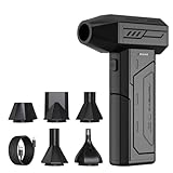
- Powerful Turbo Fan:CAGIWIRU Electric Air Duster have reached speed up to 130000RPM,Can efficient remove dust and debris.With LED light and Three Adjustable Speed to meet with different cleaning tasks.
- With Attached USB C Fasting Charging cable.Charing 3 hours,Enjoy up to 240 minutes of use on the lowest setting, For third speed can use for 25mins.with four charging options to meet with your needs.
- Wide Application: attached 5 different nozzles,making it suitable for a variety of scenes, such as car,pet, pc, keyboards, and other electronic devices. It also serves for office and home clean duster.
- Handy Design & Portable:Cordless air duster electric nozzle is Light weight and small size, design and comfortable to hold and space saving, convenient to carry around. You can put it in your room or inside the car without taking up space.
- Economical &Reusable: Built durable materials with a long-lasting battery,our CAGIWIRU air duster it's a sustainable alternative to disposable air cans.
How the reconstruction recovers depth and chemistry
Phase retrieval and ptychographic overlap
Ptychography records diffraction from a coherent probe at many positions that overlap on the specimen. Each position loses phase information in the detector measurement, but the shared regions between neighboring positions impose consistency conditions. Iterative phase-retrieval algorithms use that redundancy to recover the complex transmission function, including amplitude and phase, instead of treating each probe position as an isolated image.
Ptychographic X-ray computed tomography (PXCT) adds projections at different rotation angles. The recovered complex projection data are combined into a volumetric reconstruction. A multislice ptychographic model can represent depth-dependent propagation within a thick sample, so some depth information is obtained from the wavefield itself. That extra constraint can reduce the number of angular views needed compared with reconstructing every projection independently and then applying standard tomography.
Fluorescence for element-specific maps
Structural ptychography primarily reports complex transmission, phase and electron-density-related information. It does not automatically identify an element or chemical species. Chemical specificity comes from an energy-sensitive interaction, most directly X-ray fluorescence (XRF). When an incident pattern excites characteristic fluorescence, the detector’s energy channels indicate which elements emitted and the intensity carries information about their abundance.
Rank #2

- Screen Cleaner is Your Screen’s New Bestfriend! - EVEO is proud to present a screen cleaner that’s perfectly suited for your TV. Meet your new screen cleaner, computer screen cleaner, laptop cleaner, iPad cleaner, Macbook,tv cleaner or any other electronic device. Our screen cleaner spray and wipe along with the included plush microfiber cloth, easily removes dust, fingerprints & other smudges on the screen’s surface. Clean effectively for a streak-free screen
- Gentle on your Screens - Our screen cleaner spray and wipes is compatible to be used even for the most sensitive LCD, LED, CRT, and OLED screens in proper use. Be at ease knowing that this product is compatible with all the major brands: LG, SONY, Samsung, Sharp, iPad, iPhone, Android screens, Kindle, PC monitors - we’ve got you covered! Can be used as LCD screen cleaner, tv cleaner, smart tv screen cleaner, and more
- Premium Super Soft Plush Microfiber Cloth - Included in this efficient screen cleaning kit. This screen cleaner spray and wipe has our signature plush microfiber cloth specially designed to comply with screens and monitors and leave them streak-free. Has been shown to effectively absorbs stubborn stains, zero streaks, and no fiber shedding. Always use this together with our screen cleaner for the best results
- Easy-to-Use Screen Cleaner Kit + microfiber cleaning cloth - The EVEO Screen Cleaning Kit will easily be your new favorite tool when it comes to taking care of the things you love most, quick and easy solution for a streak-free screen. Simply screw on the sprayer gun on the screen cleaner bottle. Spray the cleaning liquid onto the microfiber cloth, then wipe the screen until desired cleanliness & shine is achieved.
- Quality Screen Care Kit Worth Purchasing - Enjoy a convenient Screen Cleaner anytime, anywhere you need it. Your satisfaction is our top priority. We stand behind the quality of our products and that’s why, you shouldn’t be worrying at all. We manufacture the Best Screen Cleaner. and can be used for laptop cleaning kit, laptop screen cleaner, tv screen cleaner for smart tv. MacBook screen cleaner, monitor cleaner or screen cleaner spray for electronic devices with microfiber cleaning cloth
In full-field structured-illumination chemical imaging, a transmission X-ray microscope projects changing patterns while an XRF detector collects an integrated signal. The correspondence between each known pattern and the measured fluorescence constrains the locations of the emitting elements. A generative reconstruction model can then estimate an elemental or compositional map without focusing the beam to nanoscale dimensions or rastering every pixel. The NxSCI demonstration retrieved compositional variations in a battery made from mixed cathode particles.
Coded-aperture dark-field depth encoding
Dark-field X-ray microscopy measures diffraction from crystalline material rather than ordinary transmission. In the coded-aperture approach, an absorbing pattern moves through the beam and casts a known silhouette across a crystalline grain. Detector pixels receive linear combinations of diffracting elements selected by that silhouette. Inverting the series of aperture positions recovers the grain’s three-dimensional structure along the diffraction-sensitive axis.
The published method reports depth-resolved grain imaging without conventional sample rotation or raster scanning. That is a different route to 3D information from PXCT: the coded silhouette supplies structured depth encoding, while the diffraction signal supplies crystallographic contrast.
Rank #3

- 【Large size】:Measuring 800 x 300 mm/ 31. 5×12 inches, which is wider and taller than others. Large mouse pad can perfectly fit your keyboard and mouse of any size, and there is also ample space for peripherals such as phones, tablets, etc.
- 【LED backlight mouse pad】:Blade Hawks RGB mouse pad provides 7 static modes and 7 dynamic modes will give you the options you are looking for. Power-off memory function remembers the last lighting mode you selected. The super glow fiber Chroma will give you a colorful gaming setup that you want.
- 【Anti-slip rubber base】:Made from Natural rubber, this gaming mousepad will firmly grip your desktop, allowing you to enjoy the ultimate precision in the games you are most passionate about. It also comes with a texture that enhances this ability even further.
- 【Smooth and waterproof surface】: Micro-textured cloth surface, for extremely precise mouse control and a smooth movement. When liquid splashes on the gaming mouse pad, it forms water droplets and slides down. Will not delay your work or gameplay.
- 【Plug and play】: This mouse and keyboard pad is powered by USB, plug and play, no driver required. One button control light modes. Press one time to change the lighting mode, press twice to change the brightness, press and hold about 3 seconds turn ON/OFF.
Why fewer measurements can be enough
Multiplexing
Each patterned exposure carries information from many locations at once. A set of distinct patterns acts like coded rows of a measurement matrix. If the rows are sufficiently independent and the detector has adequate signal-to-noise ratio, the inverse problem can recover more unknowns per exposure than a point measurement can.
The Tool Desk
Outbyte Driver Updater FREEFix the driver behind crashes, sound loss and screen glitchesFind Drivers →Outbyte PC Repair FREERepair Windows errors before they cause bigger problemsFix Now →Redundancy and overlap
Overlapping ptychographic probes repeatedly sample the same specimen regions. Those repeated observations constrain the otherwise ambiguous phase and probe estimates. Similar redundancy can come from using multiple pattern orientations, translations or X-ray energies.
Sparsity and compressibility
Many specimens are not arbitrary voxel arrays. A battery particle may contain a small number of phases; a circuit may contain large regions of nearly uniform material; a crystal grain may have a compact set of diffracting structures. Compressed-sensing penalties or piecewise-smooth models exploit that structure, allowing recovery from fewer measurements than the raw voxel count would suggest. The assumption must be tested: a highly textured, heterogeneous specimen can violate it and produce artifacts.
Rank #4

- 【Softer and More Comfortable】Vaydeer wrist rest has unique diamond pattern, which is the combination of softness and aesthetics. The materials of wrist rest are improved into higher quality memory foam and covered with silky smooth lycra. The computer wrist rest makes you as comfortable and cushiony as like rest your wrists on clouds.
- 【Ergonomic Wrist Saver】The wrist rests for keyboard and mouse comes with a 17.32×3.15×0.83 inch keyboard wrist pad and a 5.94×3.15×0.83 inch mouse wrist support. Based on ergonomic design, the unique concave shape is the perfect fit for your wrist joints. The wrist rest pad fits most computer keyboards and laptops, improve hand and wrist posture, release your wrist and arm stress.
- 【Non-Slip Rubber Bottom】Featuring an anti-skid silicone base on the bottom, this wrist keyboard support stays firmly in place on your desk, preventing the padding from sliding around, ensuring stable and consistent wrist support during extended computer sessions.
- 【Better Experience & Pain Relief】Our keyboard arm rest is beneficial to alleviate the soreness caused by direct contact and friction between your arm and a hard desk surface, reducing the risk of wrist fatigue or carpal tunnel. The soft texture of memory foam can evenly distribute the pressure around your wrists and provide good support with just enough give.
- 【Helpful in Multiple Scenarios】Whether you're working, studying, writing, typing, gaming, this keyboard and mouse rest combo is an essential accessory to add comfort and support to your hands and wrists. It’s also a great gift for men, women, family, friend, coworker, gamer, teacher, etc.
Joint volumetric inversion
Conventional tomography often reconstructs each angle separately, then combines the projections. A joint method fits all patterns and angles directly to one 3D volume. Shared parameters and consistency across views prevent each projection from spending measurements on ambiguities that the other views could resolve. This can lower the required number of angles, but it increases memory, optimization and validation requirements.
Learned regularization
Deep networks and generative models can supply a prior for severely undersampled data. They help choose plausible volumes when the measurements alone do not uniquely determine an answer. The benefit is greatest when the specimen resembles the examples or distribution used to establish the prior. The risk is bias: a network can suppress an unusual feature or create a plausible-looking one that the data do not support.
Free tools Windows power users keep installed
One-click scans. No signup required.
Three approaches and what each one reveals
| Approach | Primary signal | How 3D information is encoded | Typical scan requirements | Chemical specificity |
|---|---|---|---|---|
| Ptychographic X-ray computed tomography | Coherent diffraction and recovered complex transmission | Overlapping probe positions plus multiple projection angles; multislice models add propagation-based depth constraints | Translational probe scans and angular scans | Usually structural or electron-density contrast; chemical identification requires additional energy-sensitive measurements |
| Structured-illumination XRF (including NxSCI) | Element-specific fluorescence intensity | Known illumination patterns modulate where elements are excited; pattern-to-signal inversion estimates their 3D or projected distribution | Changing full-field patterns; no nanoscale focused raster is required | High, when fluorescence lines and calibration distinguish the elements or phases |
| Coded-aperture dark-field microscopy | Diffraction from crystalline domains | A moving absorbing aperture casts a known silhouette that selects diffracting elements and encodes depth | Aperture-position series; the demonstrated method avoids sample rotation and rastering | Crystallographic and strain-related contrast rather than direct elemental composition |
These methods are complementary rather than interchangeable. XRF answers where selected elements are, ptychography provides high-resolution complex structural information, and coded dark-field measurements target crystalline organization. A workflow that needs both chemistry and nanoscale morphology may combine signals or acquire separate patterned datasets.
Best Value

- 【No Drill Mounted】Cable management tray can be assembled in as little as 3 minutes or less-simply. Side clips easily clampe the edge of desk, pass through the holes on the sides, and secure excess cables with baskets. Desk cable management supports inward or outward installation to meet your needs in different scenarios, and provides you with great convenience when collecting and organizing wires.
- 【Rubber Pad Protects Desk】Wire organizer under desk built-in soft rubber pads on the clamp protect your desk from scratches, so you will not damage your furniture or office.
- 【Sturdy and Beautiful】Cord management under desk is made of high-quality carbon steel and holds about 15 lbs. The wire basket has 2 holes on each side of the tray to enable your cables to pass freely. Cable baskets are suitable for desk thickness use from 0.4" to 2.4".
- 【Space Saving & Keep Organized】Desk cable management tray holds all your power cords, outlet strips, USB hubs and computer transformers hidden from your feet, and the data cables will not enter the vacuum cleaner to keep your desk clean and tidy. Desk wire organizer is also suitable for use in the kitchen and outdoors.
- 【Reduce Safety Risks】Cable tray under desk keeps all plugs away from your kids, no more worry about tripping. Using 2 holes wire tray saves extra space, helps you easily cabling and wire protection. The mesh design is not easy to accumulate dust, but avoids safety accidents caused by messy wires.
What the reported experiments show
| Result | Qualification | What it demonstrates |
|---|---|---|
| 14.6 nm resolution on structures made with 22 nm technology | Prior PXCT demonstration by Holler and colleagues, as summarized in the APT publication | Nanoscale ptychographic tomography can resolve integrated-circuit features below the technology node used to fabricate them |
| More than two months estimated acquisition and reconstruction | Estimate for a 100 × 100 × 5 µm³ integrated-circuit volume containing about 2 × 10¹⁰ voxels under conventional scanning | Why volumetric raster tomography becomes impractical as field of view and resolution increase |
| 400 structured-illumination measurements per angle for a 2.8-million-voxel volume | 2026 direct X-ray fluorescence ghost-tomography preprint by Ben-Yehuda, Rack, Shwartz and Viganò; result is specific to that experiment | A highly multiplexed chemical reconstruction can operate with far fewer measurements than voxels |
| 43-fold reduction relative to raster scanning | Same 2026 preprint and experimental setup; not a universal speedup | The potential acquisition saving when pattern design, detector statistics and the reconstruction prior are favorable |
| Compositional variations recovered in mixed cathode particles | NxSCI experimental battery demonstration | Structured illumination plus XRF can distinguish chemical components in a realistic heterogeneous sample |
| 3D crystalline-grain structure from aperture-position images | 2025 coded-aperture dark-field study; no conventional rotation or rastering | A moving coded silhouette can provide depth information along the diffraction-sensitive axis |
The numerical reductions above are experiment-specific measurements or estimates. They should not be interpreted as guaranteed resolution, dose or speed for a different sample, detector, X-ray energy or beamline.
A practical reconstruction workflow
- Define the contrast you need. Decide whether the output is elemental concentration, phase composition, electron density, crystal orientation or another structural quantity. Choose XRF, coherent diffraction, dark-field diffraction or a combined signal accordingly.
- Choose a pattern family. Select probe translations, coded-aperture positions, random masks or periodic structured illumination. Patterns should be sufficiently diverse for the expected object and compatible with the source, optics and detector field of view.
- Calibrate the forward model. Measure pattern position and shape, beam energy, detector geometry and point-spread response. Include probe intensity, aperture transmission, fluorescence efficiency and background. Errors here are interpreted by the inversion as sample structure.
- Acquire multiplexed data. Record detector frames, energy channels and metadata for every pattern and, when required, every angle. Monitor drift, incident flux and radiation dose so that changes between exposures can be modeled rather than mistaken for chemistry.
- Preprocess without discarding physics. Correct dark current, flat field, detector dead time, bad pixels, background fluorescence and geometric distortions. Preserve uncertainty estimates; photon-counting statistics determine how strongly the reconstruction should trust each measurement.
- Run a joint inversion. Fit the measured data with a wave-propagation, fluorescence or diffraction forward model. Add only defensible constraints such as non-negativity, known support, sparsity or a validated learned prior. For ptychography, solve probe and object together when probe uncertainty is significant.
- Validate against held-out information. Reconstruct with withheld patterns or angles, vary the regularization, and inspect residuals. Compare chemical ratios with independent spectroscopy or known composition when available. A visually sharp result is not evidence of accuracy if the model can hallucinate missing structure.
- Report uncertainty and sampling. State the number of patterns, probe positions, angles, energy channels, dose, voxel size, detector conditions and reconstruction assumptions. Include failure cases and sensitivity to calibration so another facility can judge whether the result transfers.
Can these methods replace point-by-point tomography?
They can replace a raster strategy when the specimen and signal satisfy the assumptions that make multiplexing invertible. Full-field structured illumination is attractive for broad fields and chemical maps because it avoids nanoscale focusing. Coded-aperture dark-field imaging is useful when rotation is difficult or when depth along a diffraction axis is the key measurement. PXCT remains compelling when nanoscale structural resolution and quantitative phase are priorities and the sample can tolerate translational and angular scans.
Replacement is not automatic. A conventional scan provides direct local measurements that are easier to interpret and less dependent on priors. Patterned acquisition trades that simplicity for throughput: each voxel may receive less direct signal, noise correlations become important, and a calibration error can affect many locations at once. If the object is dense, chemically diverse or unlike the training data, the nominal measurement reduction may disappear because more patterns or angles are needed for a stable reconstruction.
Quick wins for a faster PC:
Repair Windows errors before they cause bigger problemsFix Now →Fix the driver behind crashes, sound loss and screen glitchesFind Drivers →Clear out junk files and repair common Windows errorsFree Scan →Resolution, dose and field-of-view trade-offs
Nanoscale ptychography obtains fine detail by combining coherent illumination, overlapping positions and high-quality diffraction data, but computational and acquisition cost rise quickly with volume. Full-field structured illumination covers more area per exposure and can integrate fluorescence efficiently, yet its resolution is limited by the microscope, pattern fidelity, detector sampling and conditioning of the inverse problem. Coded apertures add fabrication, alignment and flux-loss constraints; an absorbing mask can reduce photon throughput even as it removes mechanical scanning.
Fewer exposures do not guarantee lower dose. A multiplexed frame may require enough photons to estimate many unknown contributions, and repeated patterns can still deliver a substantial total dose. Dose reduction occurs when the desired reconstruction quality is achieved with fewer incident photons or when the specimen receives fewer high-intensity focused exposures. Radiation damage, drift and photon statistics must therefore be measured for the particular experiment.
Common failure modes and fixes
- Pattern misregistration: the measured pattern differs from the assumed one. Track stage coordinates and refine pattern shifts or probe positions during reconstruction.
- Insufficient pattern diversity: similar masks create nearly dependent rows in the measurement matrix. Add translations, orientations or energies that improve conditioning.
- Detector saturation or narrow dynamic range: strong fluorescence or diffraction peaks hide weak signals. Use exposure bracketing, attenuation or a detector configuration with suitable range.
- Drift and changing illumination: the object or beam moves during a scan. Interleave reference exposures, monitor flux and include time-dependent parameters.
- Over-aggressive undersampling: artifacts, ringing and loss of fine features appear. Increase patterns or angles, weaken the prior, and test reconstructions on withheld data.
- Model mismatch: fluorescence self-absorption, multiple scattering or thick-sample propagation is ignored. Upgrade the forward model or restrict claims to the regime it describes.
- Prior-induced bias: a learned or sparse model removes rare features or invents familiar ones. Compare several priors, inspect residuals and obtain an independent measurement of critical features.
- Computation becomes the bottleneck: joint 3D inversion can require large memory and long optimization runs. Use multiresolution initialization, distributed reconstruction and smaller validation volumes before scaling up.
What to specify when comparing a patterned method
“Fewer scans” is meaningful only when the baseline and quality target are explicit. Report the number of patterns per angle, number of angles, probe positions, detector frames and energy channels, then state the voxel count, field of view, resolution criterion, dose and reconstruction time. Distinguish a reduction in mechanical positions from a reduction in total photons or compute. Finally, identify whether the result was obtained with a physical model alone, a sparsity constraint or a learned generative prior. Those details determine whether the apparent 43-fold or similar saving is relevant to another specimen.
Quick Recap
Product prices and availability are accurate as of the date/time indicated and are subject to change. Any price and availability information displayed on Amazon at the time of purchase will apply.

