
Recommended Free Tools
iTechGuides is reader-supported. When you buy through links on our site, we may earn an affiliate commission. As an Amazon Associate I earn from qualifying purchases. Learn more
AI is more likely to automate selected radiology tasks and reshape workloads than to replace radiologists outright in the foreseeable future. No reliable evidence assigns a probability to wholesale replacement. A 2025 preprint projects fewer radiologist work hours over five years, but that is a conditional model—not a measured job-loss rate—and its authors say rising imaging demand may absorb productivity gains.
What the evidence says about replacement
The clearest quantified estimate comes from Curtis P. Langlotz’s 2025 task-based analysis, published as a medRxiv preprint. It projects a 33% reduction in radiologist hours worked over five years, with a modeled range of 14% to 49%. The estimate combines published evidence with the author’s judgment where evidence was unavailable, and PubMed identifies the preprint as not yet peer reviewed. It should be read as a scenario about hours, not a prediction that the same share of radiologists will lose their jobs.
The model’s largest projected effects come from drafting reports across modalities and delegating studies in radiography and mammography. Langlotz concludes that job loss is unlikely for the foreseeable future because the radiologist workforce is relatively static while imaging volumes continue to grow. That conclusion is conditional; the paper does not establish how productivity gains will translate into staffing in every market or specialty. Read the PubMed record for the preprint.
In a June 10, 2026 ACR Bulletin article, Christoph Wald, then chair of the American College of Radiology Board of Chancellors, likewise said imaging demand is growing at least as fast as the radiology workforce, citing work by ACR’s Commission on Human Resources and the Harvey Neiman Health Policy Institute. His article does not give a numeric growth rate or a market-by-market forecast. Wald wrote, “In my opinion, the near-term impact will not be the wholesale replacement of radiologists.” This is his professional view, not a measured consensus or guarantee. Read Wald’s ACR Bulletin article.
#1 Best Overall
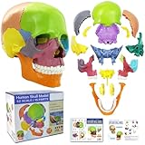
- 15 Piece Human Skull Model: This 15 parts mini human skull has every detail that the big models have, including the holes, sockets, canals, and a complete set of human teeth. Including pairs of maxilla, cheekbones, nasal bones, lacrimal bones, palatine bones and turbinate bones, and unpaired vomeres, mandibles, and hyoid bones, a total of 15 pieces, constituting the orbit and nasal cavity, bony scaffolds for the mouth and face
- Highly Detailed Anatomical Skull: It visually shows the bones structure, texture, articulating mandible, eye sockets, sutures, internal nasal cavity, and the endocranium cavity
- Easy To Reassemble: Come with an illustrated guide, complete with general knowledge and questions. This didactic skull model colored all bones (bone pairs) for easy identification and use. Individual bones can be reassembled by the inconspicuous, stable connectors mounted at the simplified suture of the skull
- Multiple Scenarios: Suitable for medical, art study, cosmetic medicine and other fields. A teaching aid for anatomy and physiology courses, display tool and lab ornament supplies
- Educational Study Tool: This human skull with brain model is suitable for neuroscience study, medical education, and professional training purposes in healthcare and anatomical sciences
Which parts of radiology AI can change
Radiology AI is not limited to reading images. The Radiological Society of North America (RSNA) describes applications in image interpretation, reporting, workflow, quantification, protocoling, education, image quality, and scan acquisition. Specific systems may assist with image classification, segmentation, measurements, object detection, triage, report-language processing, or acquisition quality. The existence of these applications does not mean every system works autonomously or is appropriate for every clinical setting. See RSNA’s statement on AI in radiology.
Report drafting and routine work
Drafting reports is a major source of projected time savings in Langlotz’s model. AI may also standardize report language and reduce avoidable variation, as Wald argues. A draft still needs appropriate review: errors in a generated report can affect downstream decisions, and performance depends on the system and context.
Rank #2

- 15 Piece Human Skull Model: This 15 parts mini human skull has every detail that the big models have, including the holes, sockets, canals, and a complete set of human teeth. Including pairs of maxilla, cheekbones, nasal bones, lacrimal bones, palatine bones and turbinate bones, and unpaired vomeres, mandibles, and hyoid bones, a total of 15 pieces, constituting the orbit and nasal cavity, bony scaffolds for the mouth and face
- Highly Detailed Anatomical Skull: It visually shows the bones structure, texture, articulating mandible, eye sockets, sutures, internal nasal cavity, and the endocranium cavity
- Easy To Reassemble: Come with an illustrated guide, complete with general knowledge and questions. This didactic skull model colored all bones (bone pairs) for easy identification and use. Individual bones can be reassembled by the inconspicuous, stable connectors mounted at the simplified suture of the skull
- Multiple Scenarios: Suitable for medical, art study, cosmetic medicine and other fields. A teaching aid for anatomy and physiology courses, display tool and lab ornament supplies
- Educational Learning Tool: This human skull with brain model is suitable for neuroscience study and comes with a color study manual to enhance the learning experience and understanding of human cranial anatomy
Triage, measurement, and workflow
Tools may flag or prioritize findings, quantify features, help route studies, or support protocol decisions. These functions can change how work is organized without transferring all interpretation or clinical responsibility to software. The practical effect depends on how much autonomy a tool has, how well it has been evaluated outside its development setting, and what happens when it fails.
Clinical synthesis and communication
Radiologists bring imaging findings together with clinical context, manage uncertainty, make recommendations, and communicate with referring clinicians and patients. Wald calls this the radiologist’s “human margin” and describes AI as an enhancer rather than a substitute. That is an attributed professional perspective, not proof that these activities can never be automated.
Rank #3

- Mini Human Skeleton Model:This scientific human model is cast from Realistically detailed miniature model of the human skeleton,are perfect for the study and clinical settings
- Size:This mini skeleton model is 17.7inches tall and with 4.75”x4.75” base.Simple and no-frills skeleton perfect for small areas
- Feature:Our mini human skeleton model s based on our US medical team,portable movement with Movable Arms and Legs,can be observed anywhere,
- Easy to stall:our anatomy desktop skeleton model is smallest and most basic ,entry-level,this mini human skeleton model is easily remove and install,a skull cap that removes from the model. The skulls jaw offers movement but does not remove from the model. The skeleton shows just the very basic anatomical detail of the spine, the ribs, and the skull.
- Excellent customer service:Our pursuit is helping student and leaner get higher level of medical study and make it easily to absorb knowledge with it
Why productivity gains do not equal job losses
If AI reduces the time needed for some tasks, the saved capacity can be used in several ways: to handle more studies, address unmet demand, reduce workload, or change staffing. Which outcome occurs depends on local imaging volume, workforce supply, adoption, and how organizations redesign work. The cited workforce analysis does not quantify those effects by region or subspecialty, so a single job-loss percentage would overstate what is known.
A 2025 AJR expert-panel narrative review identifies demand and workforce mismatch, burnout and turnover, training challenges, and AI’s potential and difficulties as issues affecting the U.S. radiologist workforce. It provides context, not a separate numeric estimate of AI-driven replacement. Read the AJR workforce review.
Rank #4

- [BUILT FOR FUTURE MEDICAL PROFESSIONALS] Designed specifically for medical, nursing, dental, PA, PT, OT, and A&P students, this life-size human skull model makes learning skull anatomy less overwhelming. With 55 numbered landmarks, a printed study card, an online PDF guide, and guided study videos, you'll build a complete learning system that makes it easier to understand anatomy, memorize structures, and prepare for lab practicals with confidence.
- [55 NUMBERED LANDMARKS FOR ACTIVE LEARNING] Instead of simply looking at a skull, actively test yourself. Identify each numbered landmark, name the structure from memory, then verify your answer using the study card or downloadable PDF. Reinforce cranial bones, sutures, foramina, and anatomical landmarks through repeated self-testing—the most effective way to retain complex anatomy.
- [REMOVABLE 3-PART DESIGN FOR TRUE SPATIAL UNDERSTANDING] Separate the magnetic skullcap and removable mandible to examine the calvaria, skull base, and lower jaw individually or together. Seeing each structure from multiple angles helps students connect textbook illustrations with real three-dimensional anatomy and better understand spatial relationships.
- [RED PAINTED SUTURES MAKE LANDMARKS EASIER TO FIND] Bright red cranial sutures contrast clearly against the bone-white PVC surface, while black numbered landmarks remain easy to read. This visual separation helps students quickly distinguish bones, sutures, and anatomical boundaries during anatomy review and practical exam preparation.
- [MOVABLE SPRING-SUPPORTED JAW FOR DENTAL & MANDIBLE STUDY] The articulated mandible opens naturally to demonstrate tooth position, occlusion, and jaw movement. Observe the relationship between the mandible and cranium during anatomy or dental training. Use gentle hand pressure to protect the spring connectors during repeated study.
What still limits safe automation
Performance may not transfer between sites
RSNA notes that many published radiology AI studies have been retrospective, conducted at one institution, and geographically limited. A tool’s performance at its development site may not carry over to another hospital or patient population. External validation matters before relying on a system in a new setting.
Performance needs ongoing monitoring
Models can change in effectiveness or degrade over time as data and workflows shift. RSNA calls for continuing monitoring and periodic recalibration; a 2024 multi-society statement also emphasizes evaluating safety and utility throughout implementation. Validation at launch is therefore not a permanent guarantee. Read the multi-society practical considerations statement.
Best Value

- This 17.72 inch full-body skeleton anatomy model restores real human skeleton proportions with clear textures of the skull, spine, ribs and joints. Highlighted with realistic details, this human skeleton model is great for medical teaching, anatomy study, art drawing, science popularization and home display.
- Designed with flexible movable arms and legs, this anatomical bone model allows free posture adjustment. Easy to assemble and disassemble, it helps you observe human structures intuitively for classroom teaching and scientific demonstration.
- Each joint features moderate damping for flexible movement and firm fixation. It can keep stable in standing, bending and other poses without loosening or shaking, ideal for learning human joint movement.
- Adopted premium eco-friendly hard PVC material, thick and sturdy. It is anti-fall, anti-crack, fade-proof and anti-yellowing, durable for long-term teaching use and daily display.
- Integrated with education and decoration, this realistic skeleton model fits for classroom, office and Halloween decor. It is also a perfect gift for students, teachers and anatomy lovers.
Generative AI can produce false information
RSNA warns that generative AI may confabulate incorrect or nonexistent information, creating potential clinical harm. Such tools need careful validation and monitoring; fluent wording alone is not evidence that a report or summary is correct.
Automation can affect human oversight
The multi-society statement discusses automation bias: clinicians may place undue trust in an automated result. It also notes that relying on automation can erode manual skill, which matters if a system fails and a clinician must take over. Safe use requires clear responsibility and workflows that preserve meaningful human review.
How widespread is radiology AI use?
A 2024 EuroAIM/EuSoMII survey contacted 28,000 European Society of Radiology members; 572 responded, a 2% response rate. Among those respondents, 274 (48%) reported currently using AI, 153 (27%) reported no use, and 145 (25%) planned to use it. Also, 255 of 572 respondents (45%) thought radiologists would take responsibility for AI outputs that may influence clinical decisions. Because participation was low, these figures describe survey respondents and should not be treated as representative of all radiologists. See the survey’s PubMed record.
The Tool Desk
Outbyte PC Repair FREERepair Windows errors before they cause bigger problemsFix Now →Outbyte Driver Updater FREEScan for outdated or missing drivers - takes under a minuteDriver Scan →What this means for radiology careers
The evidence supports preparing for changing tasks, not assuming the profession will disappear. For trainees and practicing radiologists, useful questions about an AI system or workflow include:
- Which task is affected? Distinguish drafting, triage, measurement, protocoling, and interpretation rather than treating “AI in radiology” as one capability.
- How autonomous is it? Assistance, delegated work, and autonomous decision-making carry different implications for oversight and responsibility.
- How strong is the evidence? Ask whether performance was evaluated only retrospectively at one site or externally and prospectively in relevant settings.
- How are failures handled? Look for monitoring, escalation paths, human review, and plans for recalibration.
- Where does saved time go? Productivity may meet imaging demand, reduce workload, or change staffing; the outcome is an organizational choice, not an automatic consequence of the software.
Professional guidance also points to AI literacy: clinicians need to understand a model’s intended use, limitations, and failure modes. RSNA reports that it has developed a radiology-specific AI certification program, while the ACR article stresses understanding how tools should be used. These are educational signals, not evidence that certification guarantees job security.
Quick Recap
Product prices and availability are accurate as of the date/time indicated and are subject to change. Any price and availability information displayed on Amazon at the time of purchase will apply.

